Download
1 / 41
410 likes | 529 Views
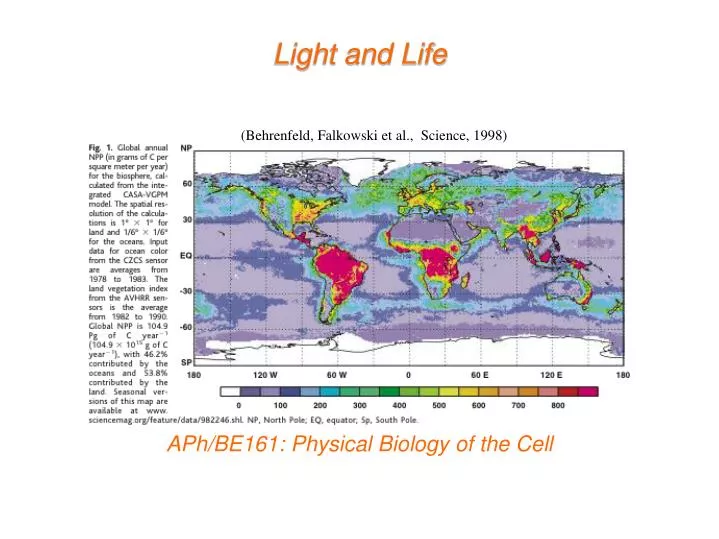
Light and Life. ( Behrenfeld , Falkowski et al., Science, 1998) . APh/BE161: Physical Biology of the Cell. What Do We Know About Eating the Sun and How Do We Know It?.

E N D
Light and Life (Behrenfeld, Falkowski et al., Science, 1998) APh/BE161: Physical Biology of the Cell
What Do We Know About Eating the Sun and How Do We Know It? The stoichiometric equation for photosynthesis tells us the key idea of the process. Energy of sunlight is converted into useful chemical bond energy in the form of sugar. Broadly speaking, the process can be conceptually divided into a part having to do with harvesting light, a part having to do with shuttling electrons and protons and a part having to do with fixing atmospheric carbon. Our plan: we will start with an overview in words and cartoons and then make some calculations on several key parts of the story.
“Nothing in Biology Makes Sense Except in the Light of Evolution” And that so includes the study of photosynthesis.
Biology’s Greatest Idea It is a great moment to reflect on evolution: 200th birthday of Darwin, 150th anniversary of his great work “On the Origin of Species” Fascinating essay of T. Dhobzhansky entitled: ``Nothing in biology makes sense except in the light of evolution.’’ - the phrase has become hackneyed, but the idea has not. Darwin found data led to inescapable conclusion, ``it was like confessing a murder’’ he wrote. Darwin’s “species question” already helps us think about the photosynthetic process.
Darwin’s Only Figure: Extinctions and Evolution The theory of evolution is built up on many different threads of evidence and one of the most important of those threads is the frequent extinctions revealed in the fossil record. Species have typical lifetimes measured in millions of years. Darwin’s one and only drawing in “On the Origin of Species” highlights extinctions.
The Great Extinctions The history of life on Earth has been punctuated by massive extinction events, some of which are famous (i.e. end of dinosaurs), but some of which are much more impressive (i.e. end Permian). For the famous dinosaur-ending extinction, a leading hypothesis argues for damage to the ability of photosynthetic organisms to collect sufficient light, with this effect propagating viciously through ecosystems. See “Extinction: Bad Genes or Bad Luck” by David Raup or “Extinction” by Doug Erwin
Losing Light and Life The leading hypothesis on the K-T extinction event is an asteroid impact in Central America though others argue for increased volcanism. One common feature in these different scenarios is a change in the light reaching Earth with a concomitant impact on photosynthetic organisms. I bring this up here as an attempt to drive home the importance of photosynthesis to life on Earth. For those with an interest in the history of Earth, photosynthesis is also a huge player in the composition of the atmosphere. http://science.nasa.gov/headlines/y2002/images/exo-atmospheres/ATM_Time_Earths.jpg
Sunlight and the Earth This curve allows us to see how the incident radiation from the sun is partitioned amongst different wavelengths. We should carry some important numbers around in our heads and one of them is 1200 W/m2 as the power available from sunlight. "The world looks so different after learning science. For example, trees are made of air, primarily. When they are burned, they go back to air, and in the flaming heat is released the flaming heat of the sun which was bound in to convert the air into tree … These things are beautiful things, and the content of science is wonderfully full of them. They are very inspiring, and they can be used to inspire others.” - Richard Feynman
Key Themes and Key Experiments The amazing story of photosynthesis passes through the conservation of matter, the discovery of the existence and nature of gases, the conservation of energy, the nature of microbes, the biochemical basis of metabolism and beyond. This subject is characterized by a long history of quantitative measurement. (Govindjee)
Gases and the Pneumochemists Van Helmont– measurement of mass change in soil during growth. He thought that the key mass transaction was the water. Ironic since he is one of the founders of the study of gases. The great “pneumochemists” – Lavoisier, Priestley, de Saussure, Ingenhousz, Senebier and others – the idea: measure the gases required for and liberated by photosyntheis. Boussingault– measured ratio of CO2taken up and O2released. Outcome: a stoichiometrically correct equation for the photosynthetic transaction that properly acknowledges the roles of water and CO2.
Light in Photosynthesis A variety of interesting experiments demonstrated that light is required for photosynthesis. Experiment of T. W. Engelmann – expose Spirogyra (challenged on wiki) (filamentous, green algae) to light of different wavelengths and see where bacteria aggregate. Answer: at places where chlorophyl absorbs. Bacteria provide a “living graph” of absorption spectrum. Starch production in leaves can be stained. Where starch is synthesized controlled by light exposure.
Light in Photosynthesis: Emerson and Arnold and Flashing Experiments A final example: clever, precision measurements of Emerson and Arnold by flashing lights of known intensity for known time periods.
The Conservation of Energy and Photosynthesis Though we all take the conservation of energy for granted, it was an idea that was hard won. Julius Robert Mayer articulated the idea and proposed that photosynthesis is a concrete example with energy from sunlight converted into chemical bond energy. Big themes that passed through photosynthesis: mass conservation, energy conservation, nature of gases, light and life.
Logic of Lecture Logical flow of lecture: we have talked about the history of our understanding of photosynthesis (very broad brush strokes) and the themes that emerged. Now, we turn to simple estimates about the overall photosynthetic productivity on the Earth and the nature of the cells that do this photosynthesis.
Use the Earth’s breathing to formulate an estimate Building a cell with photons. My daughter’s biochemistry book (and many others) tells me: every year, the earth’s plants convert 6 x 10^16 grams of carbon to organic compounds. The point of this estimate is to see how light, CO2 and H2O are used to make cells. (Behrenfeld, Falkowski et al., Science, 1998) David Keeling spent his entire career making ever more precise measurements of CO2. In his interviews, he mentions that he initially made two interesting discoveries, one of which is less famous, but extremely relevant to our story.
The Numbers: A First Look at RuBisCO and CO2 Comments on estimates, playing with numbers, sanity checks, Fermi problems, etc. How many molecules of CO2 in the atmosphere, how many carbons fixed by photosynthesis each year, how many rubisco molecules, etc.? These 1039carbons are fixed by rubisco molecules operating at a rate of roughly one carbon fixed per second (average night and day, plants and microbes).
Carbon Fixation and the Calvin Cycle Rubisco (ribulosebisphosphatecarboxylase) – enzyme that combines atmospheric CO2 with ribulose 1,5 – bisphosphate. Figure 14-40 The carbon-fixation cycle, which forms organic molecules from CO2 and H2O. The number of carbon atoms in each type of molecule is indicated in the white box. There are many intermediates between glyceraldehyde 3-phosphate and ribulose 5-phosphate, but they have been omitted here for clarity. The entry of water into the cycle is also not shown.
Calvin and Benson: “The Path of Carbon in Photosynthesis” – the role of RuBisCO The concept: where are the radioactive carbons? NOTE: as usual, when examining these classic experiments, I am struck by how blunt their instruments were and nevertheless, the reach of those discoveries. Radioactivity + chromatography. The discovery: rubisco (among other things), the machine responsible for taking atmospheric CO2 and carrying out the first steps in carbon fixation. Books claim “rubisco is the most abundant protein on earth”. Is it true? Such assertions cannot be made without some sort of justification! Let’s check the numbers. http://users.rcn.com/jkimball.ma.ultranet/BiologyPages/Chromatography_paper.html#Autoradiography
Inventories and Budgets for Some of the Most Abundant Organisms on Earth Prochlorococcus We think about the microbes that are responsible for 40% of the overall photosynthesis on Earth. Ocean water census tells us between 10,000 and 100,000 cyanobacteria per mL. This yields estimate of roughly 1026cyanobacteria doing 10% (ish) of the overall carbon fixation. Conclusion: 104rubisco per cyanobacterium. Using relatively few facts: 1 pg in 1fL with 30% of the mass ``dry’’. 30,000 Da “typical” protein tells us 3 x 10⁶ proteins. (Iancu et al, JMB, 2007)
Architecture of Cyanobacteria Every time I show you a picture of a cell, ask yourself how the architecture works. For cyanobacteria, we are going to examine several remarkable specializations related to their ability to perform photosynthesis. (Cannon et al.) (Iancu et al, JMB, 2007)
Carboxysome partitioning Very clever recent experiment that I have already told you about having to do with segregation of carboxysomes when cyanobacteria divide.
Carboxysome partitioning The carboxysome distribution can be measured. Observation: the kinds of questions they are asking are intrinsically quantitative. It doesn’t help to just look and say “there are carboxysomes”. By actually measuring the distributions, they learned something interesting about mechanism.
Carboxysome partitioning Recall your homework problem where you looked at binomial partitioning. That exact method was used here to conclude that partitioning is NOT binomial.
Photosynthetic Membranes! Prochlorococcus One of the intriguing features of these organisms is their membrane disposition. Membrane area is roughly 5 microns2. This means that the number of lipids in the outer leaflet of the bilayer is roughly 107, yielding a total of roughly 108. Membrane management: interesting and challenging. Outstanding question: what are the different volumes? Thylakoid membrane in Synechocystis (Liberton et al.)
Electron Microscopy Images of Chloroplasts We already talked about cyanobacteria. Most familiar photosynthetic organisms are plants. They have internal organelles devoted to photosynthetic process (these organelles are thought to be endosymbionts– how do we know?). Chloroplast structure is rich and fascinating, and features a complex membrane system dividing the chloroplast into three distinct spaces. Thylakoid membranes are a challenge to our understanding of biological membrane morphology.
Models of Chloroplast Structure Hierarchical description of the structure of chloroplasts. This schematic shows the three membrane-bound spaces as well as the thylakoid membrane system. Note from RP: the formation of maintenance of these membrane structures is fascinating and mysterious. NOTE: not clear how this works in cyanobacteria From Alberts, MBoC5: This photosynthetic organelle contains three distinct membranes (the outer membrane, the inner membrane, and the thylakoid membrane) that define three separate internal compartments (the intermembrane space, the stroma, and the thylakoid space). The thylakoid membrane contains all the energy-generating systems of the chloroplast, including its chlorophyll. In electron micrographs, this membrane seems to be broken up into separate units that enclose individual flattened vesicles (see Figure 14-35), but these are probably joined into a single, highly folded membrane in each chloroplast. As indicated, the individual thylakoids are interconnected, and they tend to stack to form grana.
Comparison of Mitochondria and Chloroplasts: Energy Factories of the Cell Mitochondria and chloroplasts share several interesting features. Foremost, they are both thought to be endosymbionts and have their own DNA to prove it. Complex membrane morphologies provide the seat of membrane machines responsible for ATP generation, electron transfer (and charge separation) and light harvesting (chloroplasts). From Alberts et al., MBoC5: A chloroplast is generally much larger than a mitochondrion and contains, in addition to an outer and inner membrane, a thylakoid membrane enclosing a thylakoid space. Unlike the chloroplast inner membrane, the inner mitochondrial membrane is folded into cristae to increase its surface area.
Molecules Responsible for Absorption of Light Chlorophyll characterized by a porphyrin ring and a hydrophobic tail which anchors the molecule to the membrane. The porphyrin ring is host to the electronic states that participate in the interaction with light.
Absorption Spectra of Biological Pigments The spectrophotometer permits the measurement of absorption as a function of the incident wavelength. Note that chlorophyll appears green because it absorbs strongly in the blue and the red. We will be interested in examining the quantum mechanical underpinnings of absorption spectra.
Pigments in Different Organisms Chlorophylls and other biological pigments are quite diverse (and can be used to help us classify organisms – think about it, how do you decide on evolutionary relatedness?). This table is in case you want to think about these molecules more deeply. (see “Plant Physiology” by Lincoln Taiz and Eduardo Zeiger. http://4e.plantphys.net/article.php?ch=7&id=67
How Light Energy Is Funneled Away to Perform Charge Separation: Photosystems One of the key outcomes of the Emerson-Arnold experiments was the realization that the molecular apparatus came with numbers that had an odd ratio. Two key components: 1) antenna complex and 2) photochemical reaction center. From Alberts et al., MBoC5: The antenna complex is a collector of light energy in the form of excited electrons. The energy of the excited electrons is funneled, through a series of resonance energy transfers, to a special pair of chlorophyll molecules in the photochemical reaction center. The reaction center then produces a high-energy electron that can be passed rapidly to the electron-transport chain in the thylakoid membrane, via a quinone.
Photon Flux Calculation The question: How many photons are reaching a molecule each second and what might this tell us about the nature of the photosynthetic reactions?
The Molecular Machines of Photosynthesis Figure 14-47 The structure of photosystem II in plants and cyanobacteria. The structure shown is a dimer, organized around a two fold axis (red dotted arrows). Each monomer is composed of 16 integral membrane protein subunits plus three subunits in the lumen, with a total of 36 bound chlorophylls, 7 carotenoids, two pheophytins, two hemes, two plastoquinones, and one manganese cluster in an oxygen-evolving water-splitting center. (A) The complete three-dimensional structure of the dimer. (B) Schematic of the dimer with a few central features indicated. (C) A monomer drawn to show only the non-protein molecules in the structure, thereby highlighting the protein-bound pigments and electron carriers; green structures are chlorophylls. (Adapted from K.N. Ferreira et al., Science 303:1831-1838, 2004. With permission from AAAS.)
Schematic of the Electron Transfer Process (A) The initial events in a reaction center create a charge separation. A pigment-protein complex holds a chlorophyll molecule of the special pair (blue) precisely positioned so that both a potential low-energy electron donor (orange) and a potential high-energy electron acceptor (green) are immediately available. When light energizes an electron in the chlorophyll molecule (red electron), the excited electron is immediately passed to the electron acceptor and is thereby partially stabilized. The positively charged chlorophyll molecule then quickly attracts the low-energy electron from the electron donor and returns to its resting state, creating a larger charge separation that further stabilizes the high-energy electron. These reactions require less than 10-6 second to complete. (B) In the final stage of this process, which follows the steps in (A), the photosynthetic reaction center is restored to its original resting state by acquiring a new low-energy electron and then transferring the high-energy electron derived from chlorophyll to an electron transport chain in the membrane. As will be discussed subsequently, the ultimate source of low-energy electrons for photosystem II in the chloroplast is water; as a result, light produces high-energy electrons in the thylakoid membrane from low-energy electrons in water. Schematic of the charge transfer process after optical excitation.
Measuring the Rate of Electron Transfer: the Case of Azurin Protein engineering permits construction of donor-acceptor pairs at different distances from each other. Measure the rate of electron transfer as a function of distance. (From our own Prof. Harry Gray – see his papers in PNAS)
Schematic of the Electron Transfer Process Dynamics of the electron transfer process. Are these cartoons informative? Are these cartoons “correct” and can they motivate a mathematization of the problem?
“Changes in Redox Potential During Photosynthesis” The energetics of the light-induced reactions have been worked out. The redox potential for each molecule is indicated by its position along the vertical axis. Note that photosystem II passes electrons derived from water to photosystem I. The net electron flow through the two photosystems in series is from water to NADP+, and it produces NADPH as well as ATP. The ATP is synthesized by an ATP synthase that harnesses the electrochemical proton gradient produced by the three sites of H+ activity that are highlighted in Figure 14-48. This Z scheme for ATP production is called noncyclicphotophosphorylation, to distinguish it from a cyclic scheme that utilizes only photosystem I (see the text).
The Overall Process of Photosynthesis Harvest light. Move charges. Make organic matter (sugars, starch and beyond).
Carbon Fixation and the Calvin Cycle Figure 14-40 The carbon-fixation cycle, which forms organic molecules from CO2 and H2O. The number of carbon atoms in each type of molecule is indicated in the white box. There are many intermediates between glyceraldehyde 3-phosphate and ribulose 5-phosphate, but they have been omitted here for clarity. The entry of water into the cycle is also not shown.
Different Photosynthetic Specializations Tricks to deal with low CO2 concentrations when plants close stomata. CO2 pumped into specialized cells. (A) Comparative leaf anatomy in a C3 plant and a C4 plant. The cells with green cytosol in the leaf interior contain chloroplasts that perform the normal carbon-fixation cycle. In C4 plants, the mesophyll cells are specialized for CO2 pumping rather than for carbon fixation, and they thereby create a high ratio of CO2 to O2 in the bundle-sheath cells, which are the only cells in these plants where the carbon-fixation cycle occurs. The vascular bundles carry the sucrose made in the leaf to other tissues. (B) How carbon dioxide is concentrated in bundle-sheath cells by the harnessing of ATP energy in mesophyll cells.