Download
1 / 73
730 likes | 960 Views
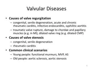
ECHOCARDIOGRAPHIC ASSESSMENT OF STENOTIC VALVULAR LESIONS. DEEPAK NANDAN. AORTIC VALVE. ANATOMY Area-2.6-3.5 cm². Structure 3 cusps,3 commissures supported by fibrous annulus Arantius nodule 3 sinuses. 2D-IMAGE. Qualitative diagnosis Thin and delicate

E N D
ECHOCARDIOGRAPHIC ASSESSMENT OF STENOTIC VALVULAR LESIONS DEEPAK NANDAN
AORTIC VALVE ANATOMY Area-2.6-3.5 cm². Structure 3 cusps,3 commissures supported by fibrous annulus Arantius nodule 3 sinuses
2D-IMAGE • Qualitative diagnosis • Thin and delicate • Plax-opening and closing • Basal short axis view-Y-inverted Mercedes Benz sign
Doppler assessment • Maximum jet velocity • BERNOULLI’s equation • Multiple windows • Parallel alignment • Colourdoppler • Angle correction
Pressure gradients-Instantaneous vMean • MIPG=4 xV²(maximal jet velocity)m/s • MPG=4x(∑V1²+V2²+…Vn²)/n • MPG=∆P(max)/1.45 +2 • MPG=2.4(Vmax)²
Bernoulli's VS invasive • Discrepancies • Tech poor doppler recording • Non parallel interrogation angle • Pressure grad depends on flow rate & valve narrowing –AR/LV dysfunction
Aortic valve area • Continuity equation:- SV (lvot)= SV (Ao) SV=CSAxTVI CSA (lvot) xTVI (lvot)=CSA (Ao) x TVI (Ao) AVA=CSA x TVI (lvot) / TVI (Ao)
Correlates well with invasive data (GORLINS) • Adv compared to Berrnoulli co-existing AR Left ventricular dysfunction
AVA-Direct planimetry • Rarely are all 3 leaflets imaged perpendicular • Triangular shape- measurement error • Deformities n irregularities- further exacerb • AV- superior-inferior rapid moments • 0.25 cm2 margin
DOBUTAMINE ECHO • Ao valve area≈Ao flow rate • Dist- true severe valvular stenosis (vs) mild to mod stenosis with LV dysfn • Stepwise infusion of dobutamine(5—30µg/kg/min)
Flexible valves:- AVA ↑ when SV ↑ • True stenotis:- AVA↔ when SV ↑ • Flexible valves:-Vmax(lvot)/jet ↑ • True stenosis:-Vmax(lvot)/jet↔ • Safe& clinically useful, limitation- non response to dobutamine
Stress findings of severe stenosis AVA<1cm² jet velocity>40m/s mean gradient>40mm of Hg • Lack of contractile reserve- failure of LVEF to ↑ by 20% is a poor prognostic sign
M- mode • Maximal aortic cusp separation (MACS) Vertical distance between right CC and non CC during systole Stenotic AV → decreased MACS • Limitations Single dimension Asymmetrical AV involvement Calcification / thickness ↓ LV systolic function ↓ CO status
OTHER APPROACHES • Ao valve resistance- flow independent measure of stenosis severity Resistance=(∆P/∆Q)mean x1333 Resistance=28√gradient( mean)/AVA
Left ventricular stroke work loss(SWL) • SWL (%) = (100 ×∆ P mean) / (∆P mean + SBP) Principle-LV expends work during systole to keep the AV open and to eject blood into the aorta Depends on the stiffness of AV Less dependent on the flow >25%--- poor outcome
Discrepencies in AS severity assessmentSevere AS by gradientSevere by area • LVOT underestimated • LVOT TVI-too far frmval • Small body size • LwtransAoflw rate low EF small vent chamber mod-sev MR mod-sev MS • LVOT overestimated • LVOT TVI recorded too close to valve • HghtransAo flow rate mod-sev AR Hgh output state Large body size
APPROACH • Valve anatomy, etiology • Exclude other LVOTO • Stenosis severity – jet velocity mean pressure gradient AVA – continuity eq • LV – dimensions/hypertrophy/EF/diastolic fn • Aorta- aortic diameter/ assess COA • AR – quantification if more than mild • MR- mechanism & severity • Pulmonary pressure
NATURAL HISTORY • Av ↑in MPG per yr = 0 to 10mm/yr mean 7mm Hg • AVA ↓ by 0.1 to ∓ 0.19cm² • Jet vel < 3m/s – rate of symptom onset needing MVR is 8 % /yr • 3-4m/s – 17%/yr • >4m/s – 40% /yr
Mitral valve-anatomy • Mitral annulus • The leaflets • Chordaetendinae-papillary muscle • Underlying ventricular wall
Leaflets • Anterior- three scallops • Posterior- three scallops • Scallop 1-lateral most • Scallop 3-medial most
Chordae and papillary muscles • Antero lateral PM- chordae to AL half of both leaflets • Dual blood supply • Postero medial PM- chordae to PM half both leaflets • RCA blood supply
2d echo-features • Maximal excursion of leaflet tips • Tubular channel
RHEUMATIC MS • Commissural fusion⇒doming/bowing • Chordal thickening ⇒ abnormal motion • Progressive fibrosis⇒stiffening ⇒calcification
Mitral stenosis 2D • Doming of the mitral valve (hockey stick AML) • Funnel shaped opening of mitral valves • Focal thickening and beading of leaflets • calcification
early diastolic doming motion of the AML, restriction of tip motion. Pliable, little fibrosis, calcification, or thickening. Dilated LA
2D short axis imaging of diastolic orifice -planimetry • Smallest orifice at the leaflet tips • Inner edge of the black/white interface traced • Correlates well with hemodynamic assessment