Download
1 / 34
390 likes | 612 Views
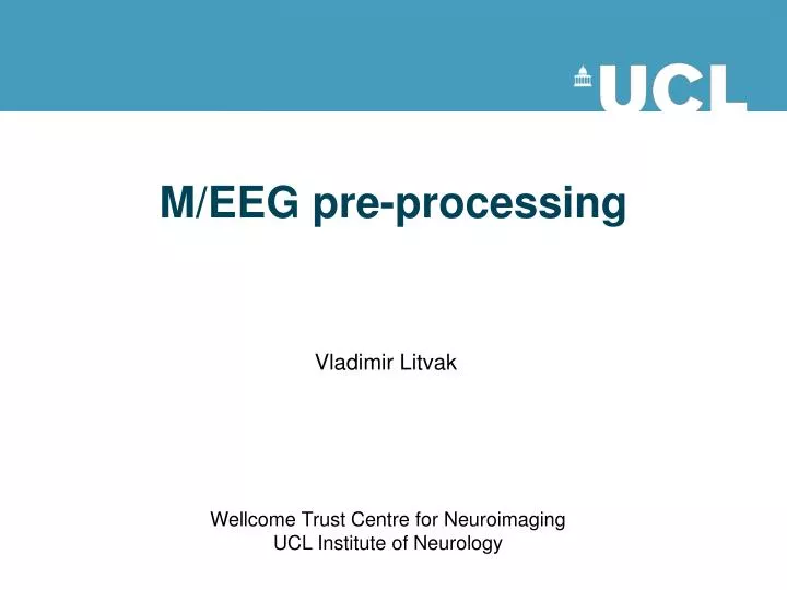
M/EEG pre-processing. Vladimir Litvak. Wellcome Trust Centre for Neuroimaging UCL Institute of Neurology. Clarification of terms – SPM speak. GUI. Script. Batch. Why batch?. As opposed to GUI - it is easy to reproduce analyses, apply them to different datasets and make modifications.

E N D
M/EEG pre-processing Vladimir Litvak Wellcome Trust Centre for Neuroimaging UCL Institute of Neurology
Clarification of terms – SPM speak GUI Script Batch
Why batch? • As opposed to GUI - it is easy to reproduce analyses, apply them to different datasets and make modifications. - removing interactive GUI elements from the code makes it cleaner and easier to maintain. • As opposed to script • complex pipelines can be built without programming expertise • saved pipelines can be examined without deciphering other people’s code • batch provides uniform interface for scripting to different parts of SPM code. • Disadvantages of batch • there is no step-by-step guiding of the user • the need to prepare all the inputs in advance makes some of the operations less intuitive and requires getting used to.
What do we need? Events Possible source of artefact M/EEG signals Time axis (sampling frequency and onset) Sensor locations Hans Berger 1873-1941
~300 sensors <128 electrodes
Evoked response vs. spontaneous activity active awake state pre-stim post-stim resting state falling asleep sleep deep sleep coma 50 uV Averaging 1 sec evoked response ongoing rhythms
Conversion MEG CTF Neuromag BTi-4D Yokogawa EEG EEGLAB Biosemi Brainvision Neuroscan EGI ANT SPM5 … Biosig Arbitrary ASCII Matlab Fieldtrip raw timelock freq GUI script Prepare (Import from workspace) + Reviewing tool Convert button (fileio) spm_eeg_convert spm_eeg_ft2spm SPM dataset *.dat – binary data file *.mat – header file
Expert’s corner • The *.mat file contains a struct, named D, which is converted to an meeg object by spm_eeg_load. • The *.dat file is memory-mapped and linked to the object. • Special functions called ‘methods’ provide a simple interface for getting information from the object and updating it and ensure that the header data remain consistent.
Sensor locations MEG: • Requires quite complex sensor representation including locations and orientations of the coils and the way MEG channels are derived from the sensors. • Sensor representation is read automatically from the original dataset at conversion. EEG: • Presently only requires electrode locations. In the future will also include a montage matrix to represent different referencing arrangements. • Usually electrode locations do not come with the EEG data. • SPM assigns default electrode locations for some common systems (extended 10-20, Biosemi, EGI – with user’s input). • Individually measured locations can be loaded; requires co-registration.
Understanding coordinate systems Coordinate systems can differ in their origin, units and orientation. • MNI coordinates are defined using landmarks inside the brain. • Advantage: locations can be related to anatomy • Disadvantage: co-registration to a structural scan is required • Head coordinates are defined based on the fiducials. Commonly used for MEG, but the definition differs between different MEG systems. • Advantage: once the location of the head is expressed in head coordinates, it can be combined with sensor locations even if the subject moves. • Disadvantage: requires fiducials; if the fiducials are moved, the coordinate system changes. • Device coordinates are defined relative to some point external to the subject and fixed with respect to the measuring device. • Advantage: head locations can be compared between different experiments and subjects. • Disadvantage: head location needs to be tracked.
Understanding coordinate systems In SPM • Before co-registration • MEG sensors are represented in head coordinates in mm. • EEG sensors can be represented in any Cartesian coordinate system. Units are transformed to mm. • After co-registration • MEG sensor representation does not change. The head model is transformed to head coordinates. • EEG sensors are transformed to MNI coordinates.
Epoching Definition: Cutting segments around events. Need to know: • What happens (event type, event value) • When it happens (time of the events) Need to define: • Segment borders • Trial type (can be different triggers => single trial type) Note: • SPM only supports fixed length trials (but there are ways to circumvent this). • The epoching function also performs baseline correction (using negative times as the baseline).
Filtering • High-pass – remove the DC offset and slow trends in the data. • Low-pass – remove high-frequency noise. Similar to smoothing. • Notch (band-stop) – remove artefacts limited in frequency, most commonly line noise and its harmonics. • Band-pass – focus on the frequency of interest and remove the rest. More suitable for relatively narrow frequency ranges.
Filtering - examples Unfiltered 5Hz high-pass 10Hz high-pass 45Hz low-pass 10Hz low-pass 20Hz low-pass
EEG – re-referencing Average reference
EEG – re-referencing • Re-referencing can be used to sensitize sensor level analysis to particular sources (at the expense of other sources). • For source reconstruction and DCM it is necessary to specify the referencing of the data. This can be done via the ‘Prepare’ tool. • Re-referencing in SPM is done by the Montage function that can apply any linear weighting to the channels and has a wider range of applications.
Artefacts Eye blink EEG MEG Planar
Artefacts • SPM has an extendable artefact detection function where plug-ins implementing different detection methods ca be applied to subsets of channels. • Presently, amplitude thresholding, jump detection and flat segment detection are implemented. • Plug-in contributions are welcome. • In addition, topography-based artefact correction method is available (in MEEGtools toolbox).
Robust averaging Kilner, unpublished Wager et al. Neuroimage, 2005
Robust averaging • Robust averaging is an iterative procedure that computes the mean, down-weights outliers, re-computes the mean etc. until convergence. • It relies on the assumption that for any channel and time point most trials are clean. • The number of trials should be sufficient to get a distribution (at least a few tens). • Robust averaging can be used either in combination with or as an alternative to trial rejection.
A note about order • There is no single correct order of steps, but here are some considerations for order choices • It is better to filter continuous data prior to epoching to avoid filter ringing artefacts in every trial. Alternatively the epochs of interest can be padded with more data and then cropped after filtering. • It is better to do high-pass filtering or baseline correction before other filtering steps to reduce filter ringing. • It is convenient to put downsampling early in the pipeline to make the subsequent steps faster. • SPM only filters channels with physiological data. So the channel types should be set correctly before filtering. • Some artefacts (e.g. discontinuous jumps or saturations) are more difficult to detect after filtering.
Fourier analysis • Joseph Fourier (1768-1830) • Any complex time series can be broken down into a series of superimposed sinusoids with different frequencies
Fourier analysis Original Different amplitude Different phase
Methods of spectral estimation – example 1 Hilbert transform Morlet 3 cycles Morlet 5 cycles Multitaper Morlet 7 cycles
Methods of spectral estimation – example 2 Morlet w. fixed window Morlet 5 cycles Optimized multitaper Hilbert transform Morlet 7 cycles Multitaper Morlet 9 cycles
Robust averaging for TF Unweighted averaging Robust averaging
TF rescaling Raw Log Rel [-7 -4]s Diff [-7 -4]s LogR [-7 -4]s Rel [-0.5 0.5]s
Thanks to: The people who contributed material to this presentation (knowingly or not): • Stefan Kiebel • Jean Daunizeau • Gareth Barnes • James Kilner • Robert Oostenveld • Arnaud Delorme • Laurence Hunt and all the members of the methods group past and present