Download

1 / 18
200 likes | 462 Views
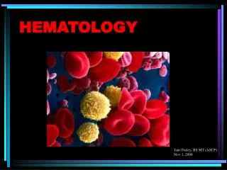
Hematology Cellcounting - Basics. Hematology, is the part of internal medicine , that is concerned with the study of blood , the blood-forming organs, and blood diseases .

E N D
HematologyCellcounting - Basics • Hematology, is the part of internal medicine,that is concerned with the study of blood, the blood-forming organs, and blood diseases. • The bunch of methods for counting, measuring and evaluating blood cells suspended in fluid is known as Hematology Cellcounting. • A blood cell analyzer, is used to perform one of medicine's most often-requested and informative diagnostic tests, the complete blood count (CBC).
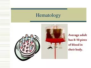
Blood Blood is a specialized fluid, „liquid tissue”, that circulatesaround the body in blood vessels. It’s main functions: - Transport through the body of • oxygen and carbon dioxide • nutrient molecules, ions, hormones • „wastes” • Heat - Defense of the body against infections, injuries and other foreign materials.
Composition of blood LiquidPlasma: over 90% of plasma is water, it also contains proteins, salts, metabolits etc. After centrifuging it is called serum. Cells: Erythrocytes(Red Blood Cells, RBC) Leukocytes(White Blood Cells, WBC) Thrombocytes(Platelets, PLT) Erythrocytes The most numerous and common type of blood cells. Non nucleated in mammals, with biconcaveshape, erythrocytes are responsible for the transport of oxygen. Contain hemoglobin(HGB), iron, and structures on the cell membranedetermine blood group systems.
Erythrocytes – cont’d Reticulocytes: immature blood cell forms, contain remnants of the nucleus(ribosomal RNA). Present normally in a very few% of the total RBC number. Hemoglobin: red blood cells contain several hundred HGB molecules which: transport oxygen(main function) contain iron cause the red colour of blood
Thrombocytes and other parameters Thrombocytes: thrombocytes or platelets play an important role in the cellular partof the hemostazis(coagulation) trying to prevent excessive blood loss. Non nucleated cells in mammals and the smallest ones inthe same time. Containspecificgranules. Hematocrit – HCT or Packed Cell Volume – PCV:Shows the volume of blood in % occupied by cells.It can be measured after separation in a centrifuge, or calculated after cell counting. Shows the degree of anemia. MeanCorpuscularVolume – MCV: Average volume of individual erythrocytes Mean Corpuscular Hemoglobin – MCH: Average hemoglobin content of erythrocytes, calculated from RBC and HGB values.MCH = HGB / RBC
Other parameters Mean Corpuscular Haemoglobin Concentration – MCHC: Calculated from the HGB and HCT values.MCHC = HGB / HCTabsolute Red cell Distribut. Width - RDW-SD Platelet Distribut. Width - PDW-SD and Red cell Distribut. Width - RDW-CV Platelet Distribut. Width - PDW-CV The distribution width of the erythrocyte or platelet population derived from the histogram at 20% of peak
Mean Platelet Volume – MPV: Average volume of individual platelets derived from the PLT histogram. Thrombocrit – PCT: Calculated from the PLT and MPV values. PCTpercentage = PLT x MPV x 100;PCTabsolute = PLT x MPV Hemolysis: rupture of the cell membrane followed by destruction of thecell. When RBC-s are lysed hemoglobin is released. Hemolytic agents: detergents, PH, osmotic pressure, drugs, immunsystem, diseases etc.
Leukocytes - WBC White blood cells are a very important part of the immune system defending the body against infectious diseases and foreign materials. By complexity of the internal structure : • Granulocytes (complex nucleus, granules in the cytoplasm) • Agranulocytes (simple nucleus, non granulated) By cell size: • „Small” • „Medium” • „Big”cells There are 5 main WBC populations: (see next page… )
Leukocytes – cont’d • Lymphocytes: mononuclear cell with round, dense nucleus and slight cytoplasm Monocytes: mononuclearcellswithoval, nucleus and variablecytoplasm (vacuoles) • Neutrophilsgranulocytes: polimorphonuclearcellswithneutralcytoplasm & granules Eosinophilgranulocytes: polimorphonuclearcellswithbasic (alkaline) cytoplasm & granules Basophilgranulocyte: polimorphonuclearcellwithacidcytoplasm & granules
Measurement principles Blood sample: whole blood mixed with EDTA, anticoagulants are necessary to prevent forming of clots
3. Hemolyser 1. 2. Blood RBC & PLT 1:32.000 WBC & HGB 1:200 Measurement principles • Sample preparation: from the whole blood a small amount(20-25ul) of primary sample is aspirated. • Diluent is added to the sample first(WBC&HGB) dilution is obtained • From this first dilution a second sample(100ul) is aspirated • Diluent is added to the second sample • RBC&PLT dilution is obtained • Lyse reagent is added to the WBC&HGB dilution RBCs are destroyed(see next slide) c b a • a + diluent → b • b + diluent → c → RBC/PLT • b + Lyser → WBC/HGB
PLT & RBC LYM MON GRA WBC differential Lysing process RBCsdestroyed WBCsselectivelyshranktonuclei
& HGB 3-part Differential WBC Histogram