Download
1 / 25
250 likes | 679 Views
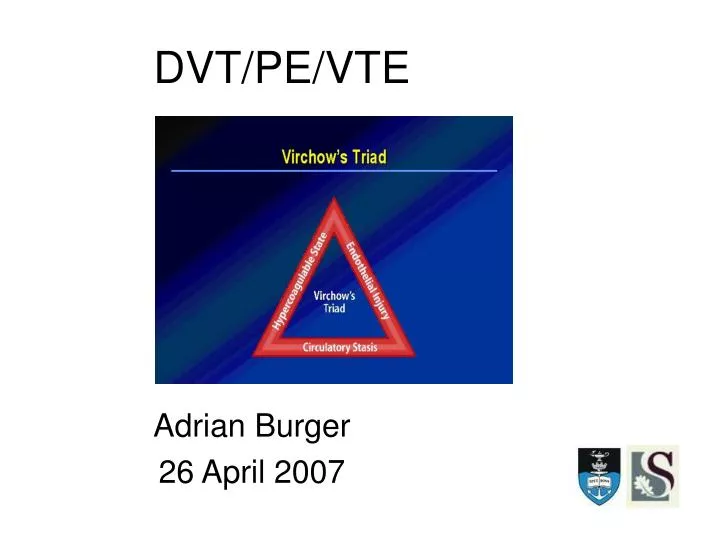
Adrian Burger 26 April 2007. DVT/PE/VTE. Virchow Triad. 3 primary components: venous stasis injury to the intima changes in the coagulation properties of the blood . Thrombus.

E N D
Adrian Burger 26 April 2007 DVT/PE/VTE
Virchow Triad 3 primary components: • venous stasis • injury to the intima • changes in the coagulation properties of the blood
Thrombus • Originates as a platelet nidus in the region of venous valves located in the veins of the lower extremities. • Further growth occurs by accretion of platelets and fibrin and progression to red fibrin thrombus, which may either break off and embolize or result in total occlusion of the vein.
Pulmonary Emboli • From the thrombi originating in the deep venous system of the lower extremities • Rarely, they may originate in the pelvic, renal, or upper extremity veins and the right heart chambers. • Large thrombi lodge at the bifurcation of the main pulmonary artery or the lobar branches, accumulate and may cause haemodynamic compromise. • Smaller thrombi continue distally, occluding smaller vessels in the lung periphery. These are more likely to produce pleuritic chest pain by initiating an inflammatory response adjacent to the parietal pleura. • Most pulmonary emboli are multiple, and the lower lobes are involved more commonly than the upper lobes
Risk Factors • age - as people over 40 are at greater risk of DVT • a past history of DVT • a family history of DVT • an inherited condition that makes the blood more likely to clot than usual • immobility • obesity • recent surgery or an injury, especially to the hips or knees • pregnancy • having recently had a baby • having cancer and its treatments • taking a contraceptive pill that contains oestrogen - but most modern pills contain a low-dose, which increases the risk by an amount that is acceptable for most women • hormone replacement therapy (HRT) - but for many women, the other benefits outweigh the increase in risk of DVT • treatment for other circulation or heart problems
Why Treat? The consequences of venous thrombosis: • Distal DVT }Symptomatic and Asymptomatic • Proximal DVT • Symptomatic PE 30% • Asymptomatic PE 40% • Fatal PE • Post-phlebitic syndrome
Wells • Low risk (-2-0) = 3-13% • Moderate risk (1-2) = 17-38% • High risk (>2)= 60-75% • Clinical Intuition = 38% underestimation
High Risk Prevalence 17-85% Moderate Risk Prevalence 0-38% Low Risk Prevalence 0-13% High Risk Prevalence 38-78% Moderate Risk Prevalence 16-28% Low Risk Prevalence 1-3% 15 DVT Studies 8 PE Studies
Clinical Decision Rule + D-dimer? • 15 Studies • Low Probability + Neg D-dimer 0.5% 3 month incidence • Moderate Probability + Neg D-dimer 3.5% 3 month incidence • High Probability + Neg D-dimer 21.4% month incidence
D-dimer Alone • Review of 5 systematic reviews Latex Turbidimetric and ELISA • Review of 78 studies ELISA & Quantitative Rapid ELISA • Cutoff 500ng/ml • Varies with age and co-morbidity • Low to Moderate Probability patients
PE ELISA 95% Sensitivity 45% Specificity Turbidimetric Assay 93% Sensitivity 51% Specificity DVT or PE ELISA 95% Sensitivity 40% Specificity Quant Rapid ELISA 97% Sensitivity 50% Specificity D-dimer Alone
Ultrasound • 8 Systematic Reviews Contrast venography as gold standard B-mode US (compression US) Duplex US – with or without colour Doppler • Symptomatic/Asymptomatic/Both • Proximal/Distal/Both
Ultrasound • Good sensitivity Symptomatic, proximal 93-100% • Good specificity all round97-100% • Poor for - asymptomatic 47-62% - upper extremity 56-100% - calf 25-93%
Ultrasound for DVT • High Sens and Spec for proximal lower extremity • Low Sens but High Spec for high risk asymptomatic patients • Poor Sens for Calf Vein • Follow-up 7 days if symptomatic and first US negative especially if moderate to high risk
CTA • 10 Systematic Reviews Old helical CT scanners PA comparison only in 4 (80-86% Sens) Sensitivities 66-93% Specificities 89-98% • At best 90% sensitivity compared with PA • Specificity better at 95%
Conclusions • Clinical Prediction Score Establish pre-test probability • D-dimer + Clinical Score High negative predictive value No further studies needed if low risk and neg • D-dimer in isolation ELISA and Rapid quantitative ELISA Young patients, short duration
Conclusion • Ultrasound Good for symptomatic, proximal lower extremity DVT Poor for asymptomatic, distal, upper extremity • CTA Newer multislice CT up to 100% sensitive
References • Ann Intern Med. 2007 Feb 6;146(3):I43. Epub 2007 Jan 29 • Ann Fam Med. 2007 Jan/Feb Vol 5 No 1 • Ann Emerg Med. 2003;42:124-135 • Emerg Med Clin North Am.Vol 22 No 3 Aug 2004