Download

1 / 19
190 likes | 347 Views
A Novel Approach to Automated Cell Counting for Studying Human Corneal Epithelial Cells. Namrata Bandekar , Alex Wong, David Clausi and Maud Gorbet. Outline. Background Problem Description Methodology Results Conclusions. Background.

E N D
A Novel Approach to Automated Cell Counting for Studying Human Corneal Epithelial Cells NamrataBandekar, Alex Wong, David Clausi and Maud Gorbet
Outline • Background • Problem Description • Methodology • Results • Conclusions
Background • Study on biocompatibility of lens cleaning solutions and lens materials • Interaction of multi-purpose solutions (MPS) with eye cells • Measure cell viability and cell activation • Human corneal epithelial cells (HCECs) acquired for in vitro study
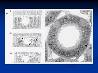
Problem Description • Identify the number of different types of cells in the grayscale images • Resolution: 1388x1040 • Scale: approximately 1 μm/pixel *Images obtained from Prof. Gorbet
Challenges • The dye fades with time • Fading time: Order of a few minutes • Cannot use global thresholding • Cell clustering • Noise • Debris • Cell shape and size • Cannot use morphology
Methodology • Detect the nuclei (local maxima) • Local thresholding • Non-maximum suppression *J. Canny, “A computational approach to edge detection,” Pattern Analysis and Machine Intelligence, IEEE Transactions on, vol. PAMI-8, no. 6, pp. 679 –698, 1986.
Methodology • Use nucleus and background seeds • Seeded region growing • Overgrows regions * R. Adams and L. Bischof, “Seeded region growing,” Pattern Analysis and Machine Intelligence, IEEE Transactions on, vol. 16, no. 6, pp. 641 –647, 1994.
Methodology • Cluster separation • Process overgrown regions • Use an adaptive thresholding technique for the clusters
Methodology • Cell body • Get seeds by considering neighbours of nuclei • Apply seeded region growing for 3 classes: background, nucleus and cell body
Testing Criterion • F1-measure where and • Accuracy * C. van Rijsbergen, Information Retrieval (2nd ed.), Butterworth-Heinemann, Newton, MA, USA, 1979.
Results • Performance compared to the distance regularized level set technique (DRLSE). • Run on a 2 GB RAM and 3.2 GHz machine. • Both algorithms implemented in MATLAB. *C. Li, C. Xu, C. Gui, and M. D. Fox, ”Distance Regularized Level Set Evolution and its Application to Image Segmentation”,IEEETransactions on Image Processing, vol. 19, no. 12, pp. 3243-3254, 2010.
Results • DRLSE fails to detect individual cells in clusters. Result of the proposed technique Original image DRLSE result
Results • DRLSE cannot detect cells with low contrast between nucleus and cell body. Original image DRLSE result Result of the proposed technique
Results • DRLSE fails to detect ghost cells. Original image DRLSE result Result of the proposed technique
Results • Performance averaged over 27 images
Results • The proposed algorithm fails in the following cases: • Overlapping ghost cells • Background illumination variation
Conclusions • Greater than 90% accuracy for nucleated cells • Robust towards low contrast • Superior to state-of-the-art techniques such as DRLSE • Future Work • Use background illumination subtraction • Concavity or notch detection for ghost cell clusters • Classification of nucleated cells in to corneal cells and white blood cells