Download
1 / 38
380 likes | 471 Views
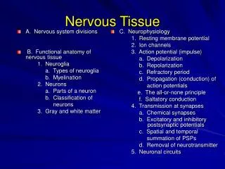
The Nervous System—made of n erves—ropelike bundles of neurons wrapped in connective tissue. Ganglia—clusters of neurons with similar functions. Central Nervous System ( CNS)= brain and spinal cord

E N D
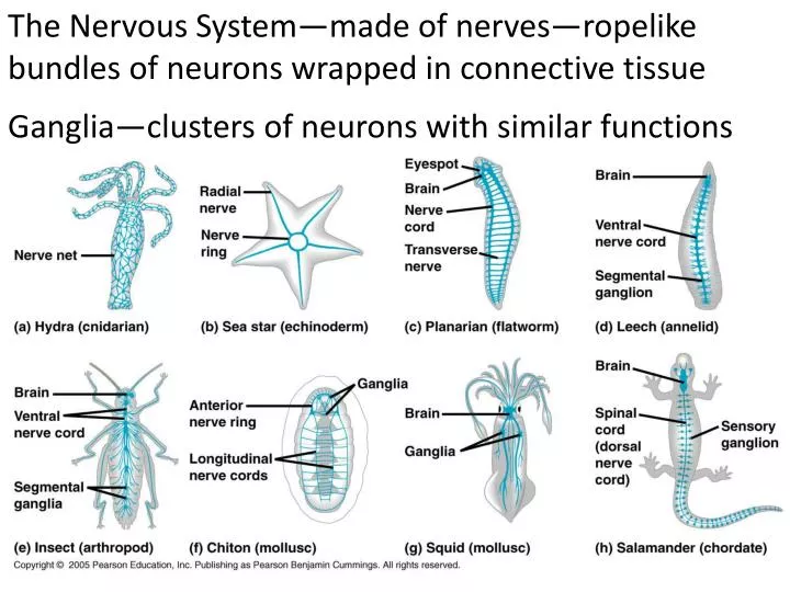
The Nervous System—made of nerves—ropelike bundles of neurons wrapped in connective tissue Ganglia—clusters of neurons with similar functions
Central Nervous System (CNS)= brain and spinal cord Peripheral Nervous System (PNS) = all the nerves BESIDES the brain and spinal cord--carry information between the CNS and the rest of the body.
Cell body—contains nucleus and organelles Dendrites—receive signals from other cells (lots/neuron) Axon—sends signals to other cells (one/neuron) Axon hillock—region of the cell body where the axon starts Myelin sheath—insulating layer around axons made up of Schwann cells (MS—deterioration of myelin sheaths) Synaptic terminals—branches at the end of the axon that send signals to other cells by releasing neurotransmitters
Synapse—site of contact between a synaptic terminal and a target cell (neuron or effector cell) Sensory neurons—send info about external and internal environments to the CNS Interneurons—intermediates between sensory neurons and motor neurons (located in the CNS) Motor neurons—send signals from the CNS to effectorcells
The knee-jerk (patellar) reflex—simple, automatic response—quadriceps contracts, flexors are inhibited from contracting
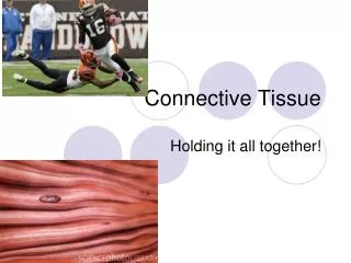
Glia—cells that support the nervous system and the functioning of neurons • Astrocytes—structural support for neurons, regulation of extracellular concentrations of ions and neurotransmitters, form tight junctions between cells lining the capillaries in the brain and spinal cord, forming the blood-brain barrier, can be stem cells
Radial glia—form tracks along which new neurons migrate, can be stem cells • Oligodendrocytes (in the CNS) and Schwann cells (in the PNS)—form myelin sheaths around the axons of vertebrate neurons during development; they wrap axons in many layers of lipid-rich membranes to provide insulation for the axons
Neurons use electrical impulses to send signals. Cells have a charge gradient across their membranes = voltage gradient The voltage measured across the membrane is the membrane potential. Usually it’s about -70 mV in an animal cell. It’s measured from the inside, so the inside of the cell has more negative ions than the outside.
The sodium/potassium ATPasepumps 3 Na+ out and 2K+in to maintain the charge gradient. Anions inside include proteins, amino acids, sulfate ions, phosphate ions. Most of these cannot cross the membrane.
Selective ion channels allow membranes to have different permeabilities to ions. The membrane potential arises from both a concentration and an electrical gradient.
The K+ ions want to go down their concentration gradient, which would be outside the cell, but that would be against their charge gradient. The membrane is more permeable to K+ than it is to Na+ Na+ wants to go in (both charge and concentration), but it goes through more slowly.
All cells have a membrane potential, but only neurons and muscle cells can change their membrane potentials. The membrane potential in the resting, unexcited state is called the “resting potential.” • Gated ion channels in the neuron membrane allow ions to move across and change the membrane potential. • Stretch-gated—open in response to mechanical stimuli • Ligand-gated—open when a neurotransmitter binds to dendrites • Voltage-gated—open when the membrane potential changes in the axon
Hyperpolarization—increasing the gradient Opening of the K+ channels would cause K+ to go OUT, making the inside of the cell MORE negative. Depolarization—reducing the gradient Opening of Na + channels would cause Na + to go IN, making the inside of the cell LESS negative. Threshold potential—if enough Na + channels open, the depolarization reaches a certain point at the axon hillock, triggering an action potential.
An action potential is an “all or none” nerve impulse. There can’t be a partial AP. • Na +channels open until threshold is reached • this is depolarization since Na +goes in, making the inside more positive • Then Na +channels close and K +channels open, sending K + this is repolarization
K+channels open and close more slowly than Na +channels do. This causes the hyperpolarization at the end of the action potential. Until the cell is back at resting potential, another action potential can’t start. This is called the REFRACTORY PERIOD. Voltage-gated channels open sequentially along the axon to propagate the action potential.
The myelin sheath insulates the axons so that there are only channels at the nodes of Ranvier…the AP jumps from node to node.
Synapses are junctions between 2 neurons, between a neuron and a muscle, or between a neuron and a gland. The presynaptic cell sends the signal. The postsynaptic cell receives the signal.
Ca++causes synaptic vesicles to release neurotransmitters that bind to receptors on postsynaptic cell membranes. NTs are quickly degraded.
EPSP = excitatory postsynaptic potential—positive ions enter the neuron in order to stimulate another action potential. IPSP = inhibitory postsynaptic potential—negative ions enter the neuron in order to inhibit an action potential. Summation: additive effect of postsynaptic potentials
Temporal summation—frequent influx of ions • Spatial summation—additive effect of many neurons signaling
Types of Neurotransmitters Acetylcholine—most common— different effects Biogenic amines—derived from amino acids—examples are epinephrine (adrenaline) and norepinephrine (they can also be hormones), dopamine, and serotonin Amino acids—GABA (inhibitory), glutamate(excitatory) Neuropeptides—ex. Endorphins Gases—made on demand and diffuse into target cells—example: nitrous oxide (NO)
Muscles—made up of fibers that are multinucleated cells. Those cells are made up of smaller fibers called myofibrils. Myofibrils are made of even smaller myofilaments. Thin myofilamentsare made of ACTIN. Thick myofilamentsare made of MYOSIN.
Each repeating unit is a SARCOMERE. Z line = sarcomere border—attached to actin I band = only actin A band = length of myosin H zone = only myosin
Sliding Filament Model Muscles contract when myosin and actin interact. Neither fiber changes in length, but they slide so that they overlap more. The I band and H zone shorten. The A band doesn’t change.
1. Myosin head bound to ATP and hydrolyzes it. 2. Myosin head binds to actin. 3. Myosin relaxes and slides. 4. Myosin binds to a new ATP to release the myosin head. Myosin is in a low-energy state when bound to ATP
Myosin binding sites on actin are blocked by tropomyosin until a neuron signals the muscle to contract. Ca++ binds to troponin, so that tropmyosin/troponin complex changes shape and exposes myosin binding sites.
Ca++ is released from the sarcoplasmic reticulum in response to an action potential. Motor neurons release acetylcholine.
Multiple motor neurons can innervate one muscle to control many fibers at once. Even though an action potential is all-or-none, a muscle contraction can be graded. If the muscle receives another AP, Ca++ levels remain high, causing a sustained contraction.
Parasympathetic: Sympathetic: “rest and digest” “fight or flight”
white matter—myelin sheaths gray matter—nerve cell bodies and dendrites
Brainstem: • Medulla oblongata--control centers for homeostatic functions (breathing, heart, digestion) • Pons—similar to medulla oblongata, conducts info between brain and spinal cord • Midbrain—receives and integrates sensory info
Cerebellum controls learning, coordination, and motor skills • Diencephalon contains • Thalamus—input & sorting center for sensory info • Hypothalamus—homeostatic regulation
Cerebrum—right and left hemispheres each with 4 lobes and connected by the corpus callosum
Cerebrum: • Planning and learning movements • Voluntary movement and cognitive functions