Download
1 / 39
400 likes | 582 Views
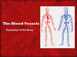
Chapter 18 Anatomy of the Blood Vessels. Introduction. The circulatory system is a series of blood vessels, or hollow tubes, that begin and end in the heart. The circulatory system delivers blood to all the body’s cells and then returns the blood to the heart.

E N D
Introduction • The circulatory system is a series of blood vessels, or hollow tubes, that begin and end in the heart. The circulatory system delivers blood to all the body’s cells and then returns the blood to the heart.
Circles, Circuits, and Circulations • The heart and blood vessels form a circle. • Two circulations: pulmonic and systemic.
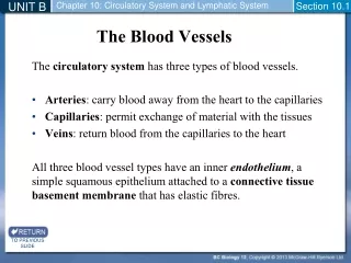
Blood Vessels • Naming the Blood Vessels • Arteries carry blood away from the heart; the smallest arteries are the arterioles. • Capillaries connect arteries and veins; a capillary is close to every cell in the body. • Veins carry blood back to the heart; small veins are called venules.
Blood Vessels - cont’d • Layers of Blood Vessels • The tunica intima is the smooth innermost layer. • The tunica media is the middle layer that contains elastic tissue and smooth muscle. • The tunica adventitia is the outermost layer of connective tissue.
Blood Vessels - cont’d • Blood Vessels: What They Do • Arteries conduct blood from the heart to the organs and are called conductance vessels. • The arterioles constrict and dilate, thereby determining resistance to the flow of blood. The arterioles are called resistance vessels. • Capillaries are concerned with the exchange of water and dissolved substances between the blood and tissue fluid. Capillaries are called exchange vessels. • Veins and venules return blood to the heart from the body. The veins also store blood and are therefore called capacitance vessels.
Major Arteries of the Systemic Circulation • The major arteries include the aorta and the arteries arising from the aorta. • See Figure 18-4 for the names and locations of the major arteries.
Major Veins of the Systemic Circulation • The major veins include the venae cavae and the veins that empty into them. • See Figure 18-5 for the names and locations of the major veins.
Special Circulations • The head and brain are supplied by two sets of arteries: the carotid arteries and the vertebral arteries. The internal carotid arteries and the basilar artery form the circle of Willis. Blood from the head and brain drains into the jugular veins. • The blood supply of the liver is composed of the portal vein, hepatic artery, and hepatic veins. The hepatic artery brings oxygen-rich blood to the liver.
Special Circulations - cont’d • The portal vein carries blood from the digestive tract to the liver. The hepatic veins carry blood from the liver to the inferior vena cava. • The fetal circulation has several unique features. The fetus uses the placenta as lungs. The umbilical blood vessels carry blood between the placenta and the fetus. Three special structures are the ductus venosus, foramen ovale, and ductus arteriosus.
The Pulse • The pulse is due to the alternating expansion and recoil of the artery creating a pressure wave (similar to vibration). • The pulse is often described as a “heartbeat that can be felt at the wrist.” • Figure 18-9 identifies the pulse points.
Introduction • The heart pumps blood through the blood vessels, supplying the cells of the body with oxygen and nutrients and carrying away the waste products of metabolism. The heart functions in a coordinated and adaptable manner to perform its tasks.
Cardiac Cycle • The cardiac cycle is a sequence of events that occurs during one heartbeat. • The events of the cardiac cycle include atrial and ventricular systole (contraction) and diastole (relaxation).
Heart: Autonomic Control • The autonomic nervous system (ANS) allows the heart to respond to changing body needs. • Stimulation of the sympathetic nerves increases heart rate (SA node), conduction velocity (AV node), and contractile force (myocardium). • Stimulation of the parasympathetic nerves (vagus) decreases heart rate and conduction velocity.
Cardiac Output (CO) • CO is the amount of blood pumped by the ventricle in 1 minute. • CO is determined by heart rate and stroke volume. • There are many factors that change HR and/or SV.
How Stroke Volume (SV) Can Be Changed • SV can be changed by Starling’s law of the heart (stretch). • SV can be changed by an inotropic effect (nonstretch).
Heart Talk • Heart Talk: includes the definition and description of commonly used clinical terms such as preload, afterload, ejection fraction, and inotropic effect.
Heart Talk - cont’d • Heart Talk: Receptor Terminology: includes the definitions of beta1-adrenergic receptor activation, beta1-adrenergic receptor blockade, muscarinic-receptor activation, and muscarinic-receptor blockade.
Left-Sided Heart Failure • The left heart can fail, producing symptoms due to a backup of blood into the pulmonic circulation (pulmonary edema). • The failing left heart is unable to pump adequate blood to the systemic circulation, producing S&S related to poor tissue oxygenation.
Right-Sided Heart Failure • Blood backs up behind the failed right ventricle, causing jugular vein distention, hepatomegaly, splenomegaly, digestive problems, and ankle edema.
Heart Failure • Heart failure is also described as backward heart failure and forward heart failure.
Introduction • The heart is a four-chambered pump that delivers blood to the lungs and the systemic circulation.
Function, Location, and Size of the Heart • The heart is located in the mediastinum toward the left side. It is about the size of a fist. • The heart pumps blood throughout the body delivering nutrients and picking up waste.
The Heart’s Layers and Covering • The heart has three layers: endocardium, myocardium, and epicardium. • The heart is supported by a slinglike pericardium. • Two layers of the pericardium form the pericardial space.
A Double Pump and Two Circulations • The right heart pumps blood to the lungs for oxygenation (called the pulmonary circulation). • The left heart pumps blood throughout the rest of the body (called the systemic circulation).
The Heart’s Chambers and Great Vessels • The heart has four chambers, two atria and two ventricles. • The atria receive the blood, and the ventricles pump the blood.
Heart Valves • The purpose of heart valves is to keep blood flowing in a forward direction. • Two atrioventricular (AV) valves are the tricuspid valve (right heart) and the bicuspid (mitral) valve (left heart). • The two semilunar valves are the pulmonic valve (right heart) and the aortic valve (left heart).
Heart Sounds • The heart sounds (“lubb-dupp”) are made by the vibrations caused by closure of the valves. • The “lubb” is due to the closure of the AV valves at the beginning of ventricular systole. The “dupp” is due to the closure of the semilunar valves at the beginning of ventricular diastole.
Pathway: Blood Flow Through the Heart • The right heart receives blood from the venae cavae and pumps it to the lungs for oxygenation. The left heart receives oxygenated blood from the lungs and pumps it to the systemic circulation. • Blood flow through the heart is summarized in the flow chart (see Figure 16-7).
Blood Supply to the Myocardium • The left and right coronary arteries supply the myocardium with oxygen and nutrients. • The coronary veins drain the unoxygenated blood and empty it into the coronary sinus (which empties into the right atrium).
Cardiac Conduction System • The heart generates an electrical signal (cardiac impulse) that moves throughout the heart in a coordinated way. The electrical signal causes the myocardium to contract. • The pathway followed by the cardiac impulse is summarized in Figure 16-10. • Cardiac muscle displays automaticity and rhythmicity. • The electrical activity of cardiac muscle is recorded as an electrocardiogram.