Download
1 / 5
50 likes | 54 Views
This text provides objectives and examples for practical classes on bone and cartilage histology. Topics include identifying different types of cartilage, understanding bone structure and cell types, and recognizing bone remodeling. Examples and questions are provided for each topic.

E N D
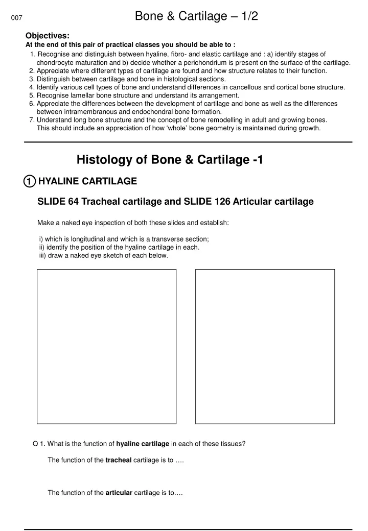
007 Bone & Cartilage – 1/2 Objectives: At the end of this pair of practical classes you should be able to : 1. Recognise and distinguish between hyaline, fibro- and elastic cartilage and : a) identify stages of chondrocyte maturation and b) decide whether a perichondrium is present on the surface of the cartilage. 2. Appreciate where different types of cartilage are found and how structure relates to their function. 3. Distinguish between cartilage and bone in histological sections. 4. Identify various cell types of bone and understand differences in cancellous and cortical bone structure. 5. Recognise lamellar bone structure and understand its arrangement. 6. Appreciate the differences between the development of cartilage and bone as well as the differences between intramembranous and endochondral bone formation. 7. Understand long bone structure and the concept of bone remodelling in adult and growing bones. This should include an appreciation of how ‘whole’ bone geometry is maintained during growth. Histology of Bone & Cartilage -1 HYALINE CARTILAGE 1 SLIDE 64 Tracheal cartilage and SLIDE 126 Articular cartilage Make a naked eye inspection of both these slides and establish: i) which is longitudinal and which is a transverse section; ii) identify the position of the hyaline cartilage in each. iii) draw a naked eye sketch of each below. Q 1. What is the function of hyaline cartilage in each of these tissues? The function of the tracheal cartilage is to …. The function of the articular cartilage is to….
Hyaline Cartilage continued Using higher magnification illustrate differences in chondrocyte shape and organisation through the depth of hyaline cartilage in each slide. Note, if present, perichondrium, young and mature chondrocytes, chondrocyte lacuna and matrix. Note the organisation and shape of the cells, particularly those actively proliferating (multiple chondrocytes in a single lacuna) and those producing new matrix (toward tissue depth). In the section of articular cartilage note the flattened appearance of chondrocytes at and close to the articulating surface. Q 2. Which cells are responsible for the synthesis of the cartilage matrix? Q 3. Name the predominant components of the extracellular matrix of hyaline cartilage? Q 4. Which sub-cellular organelles would you expect to find in cells actively involved in the synthesis of this matrix? Q 5. Does the cartilage contain blood vessels? Q 6. Did you find a perichondrium in both tissues?
ELASTIC CARTILAGE 2 SLIDE 128 Epiglottis Examine the slide at a range of magnifications and note the appearance of dense elastic fibres (stained blue to black) surrounding the chondrocytes. Sketch a small region from the centre of this cartilage. Through observation establish whether the elastic fibres have a regular or irregular arrangement and relate this to the function of the epiglottis. FIBROCARTILAGE SLIDE 130 Intervertebral disc (longitudinal section) 3 bone endplate Nucleus pulposus annulus fibrosus This series of diagrams shows the changes induced by Axial Compression and Bending. Illustrate on the RHS diagram the sites experiencing extension or compression. Examine a region as highlighted on the central figure (above). Draw a diagram illustrating the predominant orientation of concentric collagen fibre layers in the annulus. Q 7. What collagen type predominates in the Annulus Fibrosus? Q 8. Explain what you understand the term ‘slipped disc’ to mean. Check your answer! In your own time, consider the compositional similarities and differences between the three distinct types of cartilage examined.
CANCELLOUS (SPONGY or TRABECULAR) BONE SLIDE 133 Skull (Before examining slide, consider how a section through a sponge might appear!) 4 Show how the central bulk of the bone in this section might be considered to resemble your ‘section of sponge’. Select a small portion of this structure, draw an annotated diagram and identify: - Bone trabeculae - Bone marrow between trabeculae - Bone matrix - Osteocytes within the bone matrix - Osteoblasts on forming surfaces - Lining cells - Osteoclasts if present Examine the section (carefully) to identify osteoclasts. Q 9. Which of these cells is responsible for forming bone? Q 10. Which of these cells is responsible for removing bone? Q 11. Why is the cytoplasm of the osteoblast basophilic? Q 12. From which cells are osteoclasts derived and describe characteristic features of mature cells? .
5 COMPACT BONE SLIDE 131 Ground section of bone This is a transverse section through a piece of compact bone: ‘bony’ mineral alone is preserved. The spaces between this material appear black. Identify: Osteon, Concentric lamellae, Haversian canal, Osteocyte, Osteocyte lacuna, Canaliculi. Q 13. How are canaliculi formed? Q 14. Which is the predominant cell type in this section? Q 15. What does the arrangement of osteones suggest regarding Haversian remodelling? Q 16. What are Volkmann’s canals? SLIDE 132 Decalcified section of rodent bone Compare the appearance of the features seen in the ground section with those seen here. It should be noted that the bone in this section is formed from primary osteons.