Download
1 / 47
480 likes | 663 Views
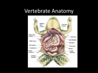
Vertebrate Tissues. OMHS Human Anatomy and Physiology Class. Tissues. Groups of cells that have specialized structural and functional roles. 4 basic types of tissues: epithelial, connective, muscle, and nervous. Classification of Tissues. Based upon shapes, arrangements, and functions:

E N D
Vertebrate Tissues OMHS Human Anatomy and Physiology Class
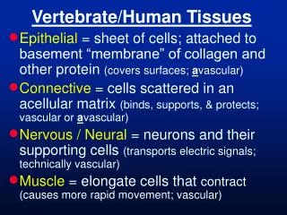
Tissues • Groups of cells that have specialized structural and functional roles. • 4 basic types of tissues: epithelial, connective, muscle, and nervous.
Classification of Tissues • Based upon shapes, arrangements, and functions: • Simple – single layer • Stratified – many layers • Cuboidal –cube shaped • Columnar – elongated shape • Squamous - flattened
A. Epithelial Tissue • Protects, secretes, absorbs. • Cover body surfaces, cover and line internal organs, compose glands. • Always has a free surface (an exposed surface).
Epithelial Cont. • Anchored to connective tissue by non-living layer called the basement membrane. • Lack blood vessels. • Readily divide (injuries heal rapidly). • Cells are tightly packed to form a good barrier.
SimpleSquamous Epithelium • Single layer of thin, flat cells. • Allows for rapid diffusion of substances; also filtration, osmosis. • Found in alveoli of lungs and lines inside of blood vessels. • Thin and delicate, easily damaged.
SimpleCuboidal Epithelium • Single layer of cube-shaped cells. • Absorption & secretion. • Lines kidney tubules, etc.
SimpleColumnar Epithelium • Single layer of tall, narrow cells. • Lines most organs of digestive tract. • Secretes digestive fluids & absorbs nutrients from food.
SimpleColumnar Epithelium Often have microvilli and secrete mucous.
Pseudostratified Ciliated Columnar Epithelium • Single row of cells- not all reach the free surface, but each cell borders the basement membrane. • Protects, secretes, & moves mucous. • Lines respiratory system - mucous traps dust, etc; cilia moves mucous out.
Pseudostratified Ciliated Columnar Epithelium Cilia Goblet Cells- secrete mucus Basement Membrane
Stratified Squamous Epithelium • Many layers of cells; cells divide in deeper layers and push older cells outward. • Layering = protection (prevents water loss and entry of chemicals, micro-organisms, etc.). • Forms epidermis; lines throat & mouth.
Stratified Squamous Epithelium Basement Membrane
Transitional epithelium • Changes in response to tension (can stretch). • Inner lining of bladder. • Protects – prevents contents of urinary tract from diffusing back into internal environment.
Glandular Epithelium • Cells that are specialized to produce and secrete substances. • Usually found within cuboidal or columnar epithelia.
4 Basic Tissue Types • Connective Tissue • Supports, binds together, protects. • Most widely distributed tissue in body. • Usually well-vascularized. • Has a matrix - material between cells • Consists of fibers and a ground substance.
(continued) • 2 types of fibers • Collagenous fibers - thick threads of protein (collagen); flexible; hold things together; white fibers. • Elastic fibers - made of protein called elastin; weaker than c.f. but stretch easily; vocal cords; yellow fibers.
1. Loose connective tissue (Areolar Tissue) • Cells are mainly fibroblasts (cells that produce fibers in the matrix). • Matrix = gel-like ground substance and many collagen and elastin fibers. • Binds skin to organs & fills space between muscles. • Has many blood vessels that nourish nearby epithelial cells.
2. Adipose Tissue (fat) • Made up of cells that store fat. • Beneath skin; between muscles; around kidneys; surface of heart; around joints. • Cushions joints and organs. • Insulates. • Stores energy.
Adipose Tissue • Large, empty-looking cells with thin margins; nucleus pressed against cell membrane.
3. Dense Connective Tissue • Densely packed, parallel collagen fibers (white) with only a few fibroblasts. • Very strong; makes up tendons and ligaments. • Low blood supply injuries slow to heal.
Dense Connective Tissue fibroblasts
4. Cartilage • Cartilage cells = chondrocytes. • Chondrocytes occupy small chambers called lacunae. a. Hyaline Cartilage • Very fine collagen fibers in matrix looks like glass. • Found on ends of bones, soft part of nose, rings that support airway, fetal skeleton.
Hyaline Cartilage lacunae Chondrocyte
b. Elastic Cartilage • Web-like mesh of elastic fibers. • Provides flexible, elastic support. • External ear and parts of larynx. Chondrocyte Lacunae
c. Fibrocartilage • Very tough, contains many collagen fibers. • Absorbs shock. • Found in meniscus of knee, intervertebral discs, etc.
Fibrocartilage lacunae chondrocyte
5. Bone • Hardness due to mineral salts and many collagen fibers in matrix. • Matrix deposited in layers called lamellae around tubes called Haversian canals. • Haversian canals contain blood vessels.
Bone (cont.) • Bone cells are called osteocytes – located in lacunae (chambers) spread out between lamellae. • Support, attachment for muscles, mineral storage, protection (cranial &thoracic cavities), forms blood cells. • Found in skeleton.
Bone Haversian Canal Osteocytes in lacunae
Bone Haversian canal Osteocyte
6. Blood • Transports materials throughout body; helps maintain homeostasis. • Matrix is fluid (called plasma).
Blood Leucocytes Thrombocytes Erythrocytes
C. Muscle Tissue • Made up of elongated cells (muscle fibers) that can contract. • Functions in movement of body parts. • 3 types: • Skeletal Muscle • Smooth Muscle • Cardiac Muscle
Skeletal Muscle • Multi-nucleated; striated – light and dark bands. • Voluntary – can be controlled by conscious effort. • Found: attached to bones.
Skeletal Muscle striations
Smooth Muscle • One nucleus; unstriated. • Found: walls of hollow internal organs- such as esophagus, intestines, stomach, blood vessels, etc. • Involuntary. • Move food through digestive tract, blood through blood vessels, etc.
Smooth Muscle Nuclei
Cardiac Muscle • Striated, one nucleus, branched. • Has intercalated disks (where cells are connected). • Involuntary. • Found only in the heart. • Pumps blood through heart chambers and into blood vessels.
Cardiac Muscle Intercalated disc
D. Nerve Tissue • Sensory reception and conduction of nerve impulses; allows for communication and coordination of body functions. • Found in brain, spinal cord, nerves. • Cells are called neurons.
Nerve Tissue Cell Body Nucleus Axon Dendrites
Essay Questions • What essay questions can you think of that would be a good assessment of your learning for this unit? • How would you………..? • What would result if…..? • Describe how…………… • Compare and contrast… • Why do you think………?