Download
1 / 41
430 likes | 1.14k Views
Lymphatic system. Lymphatic system. Plays important role in fluid distribution Important part of the immune system. Lymphatic system. Edema. Pulmonary edema. Peripheral edema. Skeletal muscle pump. Moving Blood Back to the Heart. Blood in veins is under low pressure

E N D
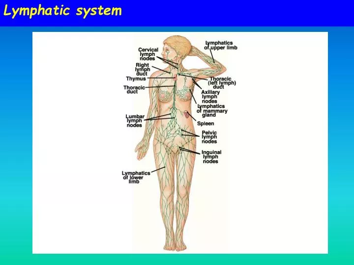
Lymphatic system • Plays important role in fluid distribution • Important part of the immune system
Edema Pulmonary edema Peripheral edema
Moving Blood Back to the Heart Blood in veins is under low pressure Two major pumps assist in moving blood back to the heart • Skeletal muscle • Respiratory pumps • Inhalation: pressure in thoracic cage drops and draws blood into veins • Exhalation: pressure increases in the thoracic cage and pushes the blood towards the heart; blood does not move backwards because of valves Figure 9.38
Veins – volume reservoir • Veins have thinner and more compliant walls • Small increases in blood pressure lead to large changes in volume • In mammals veins hold more than 60% of the blood
Regulation of circulatory systems Law of Bulk flow Q = DP/R For circulatory system CO = MAP/ TPR HR x SV = MAP/ TPR
Baroreceptor reflex • Important mechanism for regulating blood pressure • “Pressure” (stretch) receptors located in carotid and aortic bodies
Baroreceptor reflex • Baroreceptors are stretch-sensitive mechanoreceptors located in the walls of many major blood vessels • Most important of these are located in the carotid artery and aorta • Baroreceptor reflex regulates MAP Rapid changes in blood pressure are possible through sympathetic changes in cardiac output & arteriolar tone
Baroreceptor discharges • Most sensitive in physiological range • More sensitive to pulsatile pressures
Arterial Pressure and blood volume • Blood volume regulation via kidneys • Simple filtration • Humoral controls Renin-angiotensin system (RAS) Antidiuretic hormone (ADH)
Effects of gravity on blood pressure • Measured blood pressure when standing includes a hydrostatic (gravity) component • What happens when we change body positions?
How Does Gravity Affect Blood Circulation? • Very tall animals (e.g. giraffe) must be able to pump blood up to the head • They could also have difficulty with blood pooling in the feet, and peripheral edema
gill lung lung pulmonaryartery pulmonaryartery ventralaorta aorta aorta dorsalaorta Differences in central arterial blood pressures systole/diastole Human 120/80 Elephant 120/70 Pigeon 135/100 systole/diastole Trout 45/33 systole/diastole Turtle 31/25
Composition of Blood • Primarily water containing ions and organic solutes • Blood cells • Proteins
Blood Proteins • Invertebrates: primarily respiratory pigments • Vertebrates: carrier proteins such as albumin and globulins, and proteins involved in blood clotting
Blood Cells or Hemocytes Functions: oxygen transport or storage, nutrient transport or storage, phagocytosis, immune defense, blood clotting Figure 9.44
Erythrocytes • Red blood cells are the most abundant cells in the blood of vertebrates • Contain high concentrations of respiratory pigments such as hemoglobin • Major function is the storage and transport of oxygen • Have evolved independently several times
Vertebrate Blood Separates into three main components when centrifuged • Plasma • Erythrocytes • Other blood cells and clotting cells Hematocrit – fraction of blood made up of erythrocytes Figure 9.45
Erythrocytes Round or oval in shape Mammalian erythrocytes are biconcave disks • This shape increases surface area, facilitating oxygen transfer
Leukocytes • White blood cells • Function in the immune response • All are nucleated • Found both in the blood and the interstitial fluid • Some are able to move across capillary walls • Five major types Figure 9.46
Thrombocytes Play a key role in blood clotting Spindle-shaped cells in nonmammals and classified as leukocytes Anucleated cell fragments in mammals called platelets Three steps in blood clotting • Vasoconstriction • Platelet plug formation • Clot formation through coagulation cascade
Blood Cell Formation Process is called hematopoiesis Location of stem cells • Adult mammals: only in bone marrow • Fishes: kidney • Amphibians, reptiles, birds: spleen, liver, kidney, bone marrow Specific signaling factors are involved, e.g., erythropoietin is a hormone released by the kidney in response to low blood oxygen
Blood flow distribution & circulatory patterns Evolution of air breathing: Plumbing & pressures • Separation of respiratory & systemic circulations Air-breathing fishes Lungfishes Amphibians Reptiles • Mammalian fetal circulation In-series circuits 1 atrium + 1 ventricle Single pump In-parallel circuits 2 atria + 1 ventricle Single pump or afunctional double pump In-series circuits 2 atria + 2 ventricles Double pump
Fish Heart has four chambers arranged in series
Air-Breathing Fishes • Air-breathing evolved several times in fishes • Air-breathing organs are arranged in parallel; oxygenated and deoxygenated blood mix • Lungfish are the most specialized air-breathing fishes and have only limited mixing of the oxygenated and deoxygenated blood • Various organs serve as ABOs in fish • Can use: Swimbladder, mouth, gut, gills, skin, etc. • Common problems: O2 blood enters venous system mixed venous blood Single atrium receives mixed venous blood
Lungfish • Lungfish: true lung (developmentally) • Largely solved the problem of mixed venous blood • Atrium has a partial septum: oxygenated on one side; deoxygenated on the other • Ventricle has partial septum: separation maintained • Outflow vessel has a partial septum: separation is maintained
Amphibians and Reptiles • Like lungfish, the heart is only partially divided; two atria and one ventricle • Oxygenated and deoxygenated blood can mix • Two streams of blood are kept fairly separate although the mechanism is not completely understood • Crocodilians have a completely divided ventricle
Amphibians heart Anatomically undivided heart, in-parallel circulations pulmonary capillaries systemic capillaries • Single ventricle • Blood mixing • Single outflow vessel • High lung pressure • Two atria • Separate pulmonary & systemic venous return The beauty vagal vasoconstriction can closeoff lung circulationduring breath-hold or diving • Solutions • Trabecular heart • Spiral valve • Lymph hearts
Amphibian heart • Spongy myocardium & spiral valve in conus help separate oxygenated & deoxygenated blood within the single ventricle • Three chambered heart: two atria and one ventricle • Trabeculae within the ventricle and a spiral fold within the conusarteriosus help to keep oxygenated and deoxygenated blood separate
Reptilian hearts: 3 major strategies 1. Anatomically undivided heart, in-parallel circulations Squamate lizards, turtles, tortoises & most snakes • Spongy myocardium & ventricular septa (creating cava) helps separate oxygenated & deoxygenated blood within the single ventricle • Right atrium largely to cavum pulmonale • Two outflow vessels: pulmonary & systemic arteries • Blood pressures in cavum venosum, cavum pulmonale & cavum arteriosum are identical during ventricular contraction • Pulmonary capillaries protected by outflow resistance site (vagal constriction)
Reptilian hearts: 3 major strategies 2. Functionally divided heart, in-parallel circulations Varanid lizards & pythons • Highly developed ventricular muscular ridge • Systolic pressure in cavum pulmonale is lower than cavum arteriosum, but no difference during diastole: low pulmonary pressure • = functional, but not anatomical separation
Crocodilian heart: best of both worlds: 3. Anatomically divided heart, double circulation with a by-pass heart pulmonary capillaries heart systemic capillaries Right ventricle has pulmonary artery & a right systemic aorta Left ventricle has a “left” systemic aorta Foramen of Panizza provides a pressureconnection between “left” & “right” aortae Cog valve Can close off lung circulation(vagal vasoconstriction) Powered by2 atria & 2 ventricles with same wall thickness
Reptiles Can shunt blood to bypass either the pulmonary or systemic circuit • Right-to-left shunt: deoxygenated blood bypasses the pulmonary circuit and reenters the systemic circuit • Left-to-right shunt: some pulmonary blood reenters the pulmonary circuit Figure 9.16a
Mammalian fetal circulation • No air ventilation of lungs • O2 comes from placenta placenta In-series circuits 2 atria + 2 ventricles Double pump Low pulmonary pressure