Download
1 / 39
400 likes | 512 Views
Chromosomes. Cytogenetics. A subdiscipline within genetics Focuses on chromosome variations Abnormal number of copies of genes or chromosomes can lead to genetic abnormalities Human genome sequence information is used to identify genes that contribute to the chromosome-related syndromes.

E N D
Cytogenetics • A subdiscipline within genetics • Focuses on chromosome variations • Abnormal number of copies of genes or chromosomes can lead to genetic abnormalities • Human genome sequence information is used to identify genes that contribute to the chromosome-related syndromes
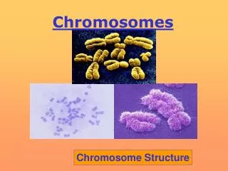
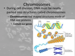
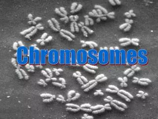
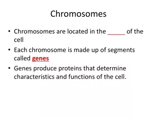
Portrait of a Chromosome • Primarily DNA and protein • Described by size and shape • Heterochromatin (dark) • Euchromatin (light) • Contains: • Telomeres • Origin of replication sites • Centromere
Chromosomes • Heterochromatin is darkly staining, contains mostly repetitive DNA • Euchromatin contains more protein encoding genes • Telomeres are chromosome tips composed of many repeats of TTAGGG and shorten with each cell division • Centromere is the largest constriction of the chromosome and where spindle fibers attach
Centromere Position • At tip telocentric • Close to end acrocentric • Displaced from center submetacentric • Long arm q • Short arm p • At midpoint metacentric
Karyotype • Chromosomal chart • Chromosomes arranged by size and structure • Arranged by largest to smallest
Visualizing Chromosomes Fetal tissue: amniocentesis chorionic villi sampling fetal cell sorting Adult tissue: blood (white blood cells) cheek swab (buccal cells) skin cells tissue biopsy
Amniocentesis Figure 13.5a
Chorionic Villi Sampling Figure 13.5b
Fetal Cell Sorting Figure 13.5c
FISHing • Fluorescence in situ hybridization • in situ =in tissue • in vivo =in living organism • in vitro =in a dish • Uses a fluorescent probe to detect specific sequences of DNA
Abnormal Chromosome # -Polyploidy • Polyploidy • An entire extra set of chromosome • Example: Triploidy • One egg fertilized by 2 sperm OR • A diploid egg fertilized by one sperm • A diploid sperm fertilizes an egg
Abnormal Chromosome # -Aneuploidy • Missing a single chromosome (monosomy) • OR - • Having one extra chromosome (trisomy) • Euploid =“good set” • Aneuploid =“not good set” • Caused by nondisjunction during meiosis
Nondisjunction at Mitosis • Results in a mosaic • Some cell populations are affected while others are not • Severity of symptoms depends on how early in development the nondisjunction occurs.
Autosomal Aneuploids • Usually lethal • Those that survive often have mental retardation • Most common for chromosomes 13, 18, and 21. Why?
Trisomy 21 • October is Down Syndrome Awareness Month!
Oogenesis Before birth Arrested in Prophase I After puberty (each month) Arrested in Metaphase II Upon fertilization
Trisomy 18 • Edward syndrome • Severe physical and mental disabilities • Development stops at the 6 month level • Oddly clenched fists • Low-set ears • Small mouth • Unusual or absent fingerprints • Liver and heart problems
Trisomy 13 • Patau syndrome • Fusion of the eyes or a small or absent eye • Cleft lip and palate • Extra fingers and toes • Mental retardation
Sex Chromosome Aneuploids: Female • Turner syndrome (XO) • Delayed puberty • 99% are not born • Infertile • Triplo-X (XXX) • Tall stature • Menstrual abnormalities • All but 1 X is inactivated
Sex Chromosome Aneuploids: Male • Klinefelter Syndrome (XXY) • Sexually underdeveloped • Small testes • Sparse facial and pubic hair • Long arms and legs and big feet and hands • May develop breast tissue • Often infertile
Sex Chromosome Aneuploids: Male • XXYY Syndrome • Slightly delayed childhood development • Behavioral problems • ADD, OCD, and learning disabilities • Leg ulcers due to poor circulation • Sexual development is delayed • Testes do not descend • Infertile • Abnormal (YY) sperm AND abnormal (XX) egg
Sex Chromosome Aneuploids: Male • Jacobs Syndrome (XYY) • 1/1000 males has an extra Y • 96% of XYY males are normal • Tall height and acne • Criminals with chromosomal abnormalities tend to have XYY
Abnormal Chromosome Structure • Deletion =missing genetic material • Can range in size (the more genes deleted, the worse the phenotype) • Duplication =a region of the chromosome where genes are repeated • Inversion =the DNA sequence in a region of the chromosome is inverted • Translocation =a piece of the chromosome is moved to another chromosome
Translocation • Nonhomologous chromosome exchange segments Two major types: • Robertsonian translocation • Two nonhomologous acrocentric chromosomes break at the centromere and long arms fuse. The short arms are often lost. • 5% of Down syndrome results from a Robertsonian translocation between chr 21 and chr 14. • Reciprocal translocation • Two nonhomologous chromosomes exchange a portion of their chromosome arms.
Reciprocal Translocation • Exchange of material from one chromosome arm to another • Some individuals carry a translocation but are not missing any genetic material unless a translocation breakpoint interrupts a gene
Inversions • Inverted chromosomes have a region flipped in orientation • 5-10% cause health problems probably due to disruption of genes at the breakpoints • Inversions may impact meiotic segregation • Two types of inversions occur: • Paracentric • inverted region does NOT include centromere • Pericentric • inverted region includes centromere
Segregation of a Paracentric Inversion Figure 13.21
Segregation of a Pericentric Inversion Figure 13.22
Isochromosomes • Chromosomes with identical arms • Form when centromeres divide along the incorrect plane during meiosis Figure 13.23
Ring Chromosomes • Chromosomes shaped like a ring • Occur in 1 in 25,000 conceptions • May arise when telomeres are lost and sticky chromosome end fuse • Radiation exposure