Download
1 / 57
570 likes | 575 Views
Explore the five special senses and their organs, including vision, hearing, smell, taste, and touch. Learn about taste buds, olfactory receptors, the structure of the eye, and more.

E N D
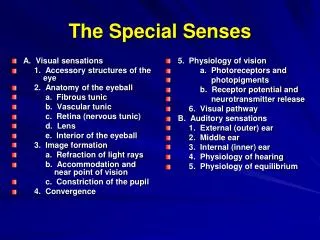
Five Senses • Vision (Sight) • Hearing • Smell • Taste • Touch (Palpation)
Taste Buds • Most of the 10,000 or so taste buds are found on the tongue • Taste buds are found in papillae of the tongue mucosa • Papillae come in three types: filiform, fungiform, and circumvallate • Fungiform and circumvallate papillae contain taste buds
Taste Buds Figure 15.1
Taste Sensations • There are five basic taste sensations • Sweet – sugars, saccharin, alcohol, and some amino acids • Salt – metal ions • Sour – hydrogen ions • Bitter – alkaloids such as quinine and nicotine • Umami – elicited by the amino acid glutamate
Sense of Smell • The organ of smell is the olfactory epithelium, which covers the superior nasal concha • Olfactory receptor cells are bipolar neurons with radiating olfactory cilia • Olfactory receptors are surrounded and cushioned by supporting cells • Basal cells lie at the base of the epithelium
Sense of Smell Figure 15.3
Vision • 70% of all sensory receptors are in the eye • Most of the eye is protected by a cushion of fat and the bony orbit • Accessory structures include eyebrows, eyelids, conjunctiva, lacrimal apparatus, and extrinsic eye muscles
Eyebrows • Coarse hairs that overlie the supraorbital margins • Functions include: • Shading the eye • Preventing perspiration from reaching the eye • Orbicularis muscle – depresses the eyebrows • Corrugator muscles – move the eyebrows medially
Palpebrae (Eyelids) • Protect the eye anteriorly • Palpebral fissure – separates eyelids • Canthi – medial and lateral angles (commissures)
Palpebrae (Eyelids) • Lacrimal caruncle – contains glands that secrete a whitish, oily secretion (Sandman’s eye sand) • Tarsal plates of connective tissue support the eyelids internally • Levator palpebrae superioris – gives the upper eyelid mobility
Palpebrae (Eyelids) • Eyelashes • Project from the free margin of each eyelid • Initiate reflex blinking • Lubricating glands associated with the eyelids • Meibomian glands and sebaceous glands • Ciliary glands lie between the hair follicles
Palpebrae (Eyelids) Figure 15.5b
Conjunctiva • Transparent membrane that: • Lines the eyelids as the palpebral conjunctiva • Covers the whites of the eyes as the ocular conjunctiva • Lubricates and protects the eye
Lacrimal Apparatus • Consists of the lacrimal gland and associated ducts • Lacrimal glands secrete tears • Tears • Contain mucus, antibodies, and lysozyme • Enter the eye via superolateral excretory ducts • Exit the eye medially via the lacrimal punctum • Drain into the nasolacrimal duct
Lacrimal Apparatus Figure 15.6
Extrinsic Eye Muscles • Six straplike extrinsic eye muscles • Enable the eye to follow moving objects • Maintain the shape of the eyeball • Four rectus muscles originate from the annular ring • Two oblique muscles move the eye in the vertical plane
Summary of Cranial Nerves and Muscle Actions • Names, actions, and cranial nerve innervation of the extrinsic eye muscles
Structure of the Eyeball Figure 15.8a
Fibrous Tunic • Forms the outermost coat of the eye and is composed of: • Opaque sclera (posteriorly) • Clear cornea (anteriorly) • The sclera protects the eye and anchors extrinsic muscles • The cornea lets light enter the eye
Vascular Tunic (Uvea) • Has three regions: choroid, ciliary body, and iris • Choroid region • A dark brown membrane that forms the posterior portion of the uvea • Supplies blood to all eye tunics
Vascular Tunic • A thickened ring of tissue surrounding the lens • Composed of smooth muscle bundles (ciliary muscles) • Anchors the suspensory ligament that holds the lens in place Ciliary Body
Vascular Tunic Iris • The colored part of the eye • Pupil – central opening of the iris • Regulates the amount of light entering the eye during: • Close vision and bright light – pupils constrict • Distant vision and dim light – pupils dilate • Changes in emotional state – pupils dilate when the subject matter is appealing or requires problem-solving skills
Pupil Dilation and Constriction Figure 15.9
Sensory Tunic: Retina • A delicate two-layered membrane • Pigmented layer – the outer layer that absorbs light and prevents its scattering • Neural layer, which contains: • Photoreceptors that transduce light energy • Bipolar cells and ganglion cells • Amacrine and horizontal cells
Sensory Tunic: Retina Figure 15.10a
The Retina: Ganglion Cells and the Optic Disc • Ganglion cell axons: • Run along the inner surface of the retina • Leave the eye as the optic nerve • The optic disc: • Is the site where the optic nerve leaves the eye • Lacks photoreceptors (the blind spot)
The Retina: Ganglion Cells and the Optic Disc Figure 15.10b
The Retina: Photoreceptors • Rods: • Respond to dim light • Are used for peripheral vision • Cones: • Respond to bright light • Have high-acuity color vision • Are found in the macula lutea • Are concentrated in the fovea centralis
Blood Supply to the Retina • The neural retina receives its blood supply from two sources • The outer third receives its blood from the choroid • The inner two-thirds is served by the central artery and vein • Small vessels radiate out from the optic disc and can be seen with an ophthalmoscope
Inner Chambers and Fluids • The lens separates the internal eye into anterior andposterior segments • The posterior segment is filled with a clear gel called vitreous humor that: • Transmits light • Supports the posterior surface of the lens • Holds the neural retina firmly against the pigmented layer • Contributes to intraocular pressure
Anterior Segment • Composed of two chambers • Anterior – between the cornea and the iris • Posterior – between the iris and the lens • Aqueous humor • A plasmalike fluid that fills the anterior segment • Drains via the canal of Schlemm • Supports, nourishes, and removes wastes
Anterior Segment Figure 15.12
Refraction and Lenses • When light passes from one transparent medium to another its speed changes and it refracts (bends) • Light passing through a convex lens (as in the eye) is bent so that the rays converge to a focal point • When a convex lens forms an image, the image is upside down and reversed right to left
Refraction and Lenses Figure 15.16
Photoreception: Functional Anatomy of Photoreceptors • Photoreception – process by which the eye detects light energy • Rods and cones contain visual pigments (photopigments) • Arranged in a stack of disklike infoldings of the plasma membrane that change shape as they absorb light
Photoreception: Functional Anatomy of Photoreceptors Figure 15.19
Rods • Functional characteristics • Sensitive to dim light and best suited for night vision • Absorb all wavelengths of visible light • Perceived input is in gray tones only • Sum of visual input from many rods feeds into a single ganglion cell • Results in fuzzy and indistinct images
Cones • Functional characteristics • Need bright light for activation (have low sensitivity) • Have pigments that furnish a vividly colored view • Each cone synapses with a single ganglion cell • Vision is detailed and has high resolution
The Ear: Hearing and Balance • The three parts of the ear are the inner, outer, and middle ear • The outer and middle ear are involved with hearing • The inner ear functions in both hearing and equilibrium • Receptors for hearing and balance: • Respond to separate stimuli • Are activated independently
The Ear: Hearing and Balance Figure 15.25a
Outer Ear • The auricle (pinna) is composed of: • The helix (rim) • The lobule (earlobe) • External auditory canal • Short, curved tube filled with ceruminous glands
Outer Ear • Tympanic membrane (eardrum) • Thin connective tissue membrane that vibrates in response to sound • Transfers sound energy to the middle ear ossicles • Boundary between outer and middle ears
Middle Ear (Tympanic Cavity) • A small, air-filled, mucosa-lined cavity • Flanked laterally by the eardrum • Flanked medially by the oval and round windows • Epitympanic recess – superior portion of the middle ear • Pharyngotympanic tube – connects the middle ear to the nasopharynx • Equalizes pressure in the middle ear cavity with the external air pressure
Ear Ossicles • The tympanic cavity contains three small bones: the malleus, incus, and stapes • Transmit vibratory motion of the eardrum to the oval window • Dampened by the tensor tympani and stapedius muscles
Middle Ear (Tympanic Cavity) Figure 15.25b
Inner Ear • Bony labyrinth • Tortuous channels worming their way through the temporal bone • Contains the vestibule, the cochlea, and the semicircular canals • Filled with perilymph • Membranous labyrinth • Series of membranous sacs within the bony labyrinth • Filled with a potassium-rich fluid
Inner Ear Figure 15.27