Download
1 / 51
520 likes | 685 Views
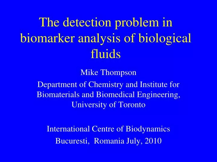
The detection problem in biomarker analysis of biological fluids. Mike Thompson Department of Chemistry and Institute for Biomaterials and Biomedical Engineering, University of Toronto International Centre of Biodynamics Bucuresti, Romania July, 2010. Clinical and Biomarker Targets.

E N D
The detection problem in biomarker analysis of biological fluids Mike Thompson Department of Chemistry and Institute for Biomaterials and Biomedical Engineering, University of Toronto International Centre of Biodynamics Bucuresti, Romania July, 2010
Clinical and Biomarker Targets Province of Ontario • 1 billion dollars annually for hospital and central lab assays (22 bd Provincial Budget) • Many assays involve magnetic bead ELISA • High level of automation but chemistry is often “old” • Virtually no introduction of lab-on-a-chip or sensor technology • Blood, urine and tissue are extremely difficult matrices
Label Free Detection Methods • Transverse wave acoustic physics in the FIA liquid-phase mode –protein small-molecule interactions, neuron cell behavior, nucleic acid damage by oxidants • Electromagnetic detection based on the propagation of ultra high frequency (1 GHz) acoustic physics • Kelvin current detection in scanning format and time-dependant measurements over nucleic acids, proteins and neurons on substrates such as ITO
Topics • Transverse wave acoustic physics as a sensor detection strategy - examples • Ultra high frequency electromagnetic physics • Model probe attachment to EMPAS surface • Linker chemistry and minimization of the pervasive NSB biosensor problem • Applications – preliminary work on the detection of ovarian cancer and HIV in serum • Application – collaboration with UK MOD • Outline of work on scanning Kelvin detection
C0 Rm Lm Cm Viscous Liquid rL, hL Liquid Biolayer Measure: fs – Energy storage Rm – Energy dissipation
Neuron culture in acoustic vibrational fields (TSM) CO2 in CO2 out growth medium and or drugs in growth medium and or drugs out Microscopic image of neurons (N-38) 2ml chamber for neuron growth Metabolic line over 48hrs
Cellular Oscillations COHERENT SYNCHRONOUS SIGNAL OF 2 MINUTES PERIODICITY!
Criteria for Protein or Aptamer Probe Attachment to Device • Molecular assembly for reproducible surface – BUT? • Si dioxide - silanization chemistry • High receptor site packing density • Capability for steric control of density • Simple bi-functionality allowing 100% reaction with probe • Minimize or eliminate NSB in biological fluids – blood, serum, urine
General Probe Model • Develop a new generation of linkers onto which thiol-containing biomolecules could immobilize in a subsequent step for the purpose of fabricating EMPAS biosensing interface • Biotin-avidin was chosen as a model system in order to test the viability of our biosensor • Chemically modified biotin to yield a thiol group on its tail
Long Alkyl Backbone Trichlorosilyl Tail Functionalizable Head Group Alkyltrichlorosilane Linkers • Trichlorosilyl tail shows strong affinity to quartz crystal • Forms a strong Si-O bond on the surface of quartz crystal • The Head function can be modified to immobilize target biomolecules
Thiosulfonate Chemistry • Thiosulfonate was chosen as the head function • Known to react chemoselectively with thiols to form disulfide bonds Gamblin, D. P.; Garnier, P.; Ward, S. J.; Oldham, N. J.; Fairbanks, A. J., and Davis, B. G. Org. Biomol. Chem. 2003, 1(21), 3642-3644.
Trichlorosilyl Undecenyl Benzene ThioSulfonate (TUBTS) • Synthesis
XPS analysis for biotinthiol immobilization on TUBTS SAMs at various time • XPS peak profile for N • N signal is unique to biotinthiol
Chemoselectivity of TUBTS SAM + N. R.
An Example of EMPAS measurement Injection of 0.1 mg/mL avidin solution (50 µL) Frequency shift of 17900 Hz
EMPAS measurements for TUBTS SAM • Specific to non-specific ratio – 1.5:1 • Acceptable reproducibility
OEG-TUBTS • Synthesis
EMPAS measurements for OEG-TUBTS SAM • Specific to non-specific ratio – 1.75:1 • High reproducibility
Incorporation of diluent • Next Step: Incorporation of a diluent molecule in our system • A diluent - a shorter molecule used to space out the linker within the SAM • Provides greater space for the analyte to interact with the biosensing element • Also attempted the biotinthiol immobilization under aqueous conditions
7-OEG • Synthesis
EMPAS measurements for OEG-TUBTS/7-OEG SAM • Specific to non-specific ratio – 2:1 • High reproducibility • Immobilization under aqueous condition is possible
OEG-TUBTS/7-OEG SAM formation on quartz crystal: Time Trial • CAM and XPS values both continued to change after 120 min • Indicated that the silanization process was not complete by 120 min
OEG-TUBTS/7-OEG SAM formation on quartz crystal: Time Trial (cont’d) • Closer look at the %F and %S in XPS analysis • Sulfur unique to OEG-TUBTS • Fluorine unique to 7-OEG • Possible multilayer formation • Dramatically decrease the biosensing performance of our surface • Decreased the silanization time to 60 min to avoid multilayer formation
EMPAS measurements for OEG-TUBTS/7-OEG SAM with reduced silanization time • Specific to non-specific ratio – 15:1 • High reproducibility
Conclusions for work on linker • Successfully prepared SAMs onto piezoelectric quartz crystals with new thiosulfonate-based linkers • Chemoselectively immobilized biotinthiol under aqueous conditions in a single, straightforward, reliable and coupling-free manner • With OEG-TUBTS/7-OEG system, we demonstrated a 15-fold difference in signal response of EMPAS between specific and non-specific measurements for avidin interaction • Same chemistry for device in goat serum spiked with avidin gives a 6-fold signal ratio – best we have ever observed
And what we have learned • Proteins adsorb to hydrophilic and hydrophobic surfaces just about equally • Modified optically flat surfaces with SAMs in place produce high NSB • For steric reasons you need a receptor functioning in tandem with a surface diluent • The linker chain length must be about 5 C longer than the diluent • PEG functionality does reduce NSB very significantly • Receptor exclusion volume plays a crucial role
Ovarian Cancer Overview • Most serious gynaecologic cancer with ≈ 1700 deaths every year in Canada • Cancer patients develop a mechanism to evade and suppress the immune system • Ovarian cancer cells have • reduced expression in signal transducing zeta chain molecules (e.g. CD3-zeta) • reduced expression of T-cell receptor molecules ( and ) • suppressed T-cell activation and proliferation • reduced cytokine production and proliferative response
Ovarian Cancer Cause • Proteomic studies revealed an early pregnancy factor (EPF) in the serum and urine of pregnant women during the 1st and 2nd trimesters • This EPF has been identified as a heat shock protein 10 (HSP10) • Cancer cells were found to produce HSP10 and release it to the cytoplasm, extracellular ascites and peripheral blood • HSP10 was associated with the reduction of T-cell CD3-zeta expression and immunosuppression
5) On-line Detection of HSP10: TSM Response Possible indication of aptamer conformational change upon HSP10 binding
Detection of HIV Antibodies in Blood • Screening test for HIV takes 3 drops of blood and effected in 2.5 minutes • Commercial kits available in several countries such as China, India and Canada • Confirmatory test for HIV requires positive detection of 10 Ab in blood • Confirmation uses electrophoresis and blotting, 3 days and is costly
Towards Multiplexed HIV Ab Detection Using Acoustic Wave Physics • Develop flow-through label-free EMPAS electromagnetic system for diagnostic assays • Attachment of probe (antigen/peptide) to device surface • Surface chemistry to maximize analytical signal and minimize response for NSB (serum-blood?) • Design engineer multiplexed system • Extend to replace ELISA approach to diagnostics
Collaboration with UK MOD Porton Down • MOD has developed rapid response SPR system for detection of bacteria/viruses • Similar to diagnostics – based on Ab/aptamer probes on gold substrate • Serious issue with interference of particles/non-specific binders • Developed long-chain, PEG thiol linker
Principle of Scanning Kelvin nanoprobe Lord Kelvin The original apparatus of Lord Kelvin
eV Evacuum 1 2 2 1 2 1 2 1 + + + + - - - - 2 1 1 + d V0= -V The Scanning Kelvin Nanoprobe is Based on the Measurement of the Local Work Function At electrical contact, equalization of Fermi levels, surface charging, electron flow Inclusion of a backing potential V0, null-field condition achieved when V0 = -V Two metals are separated by a distance d
piezo driver signal generator 2kHz Vibration piezo power supply Charge amplifier CPD signal tip lock-in amp. 1 sample insulator Topography control piezo topography signal lock-in amp. 2 XY-scantable piezo driver Sum circuit signal generator 100kHz motors control PC with LabView NI PCI 6160 DAQ Board C-842.20 DC Motor Controller NI BNC-2120 interface Sample voltage shielded cable Block Diagram of the Scanning Kelvin Nanoprobe
DNA Microarrays Array map showing the exact position of duplicates and the number of mismatches Surface potential image of the scanned oligonucleotide microarray
Protein Microarrays Image of Rabbit IgG protein microarray (35 spots in a 7x5 grid) showing the dependency of work function level on the protein abundance in different spots
Mike Thompson Research Group 2010 • Jack Sheng Sumra Bokhari • Sonia Sheikh Dr. Larisa Cheran • Shilin Cheung Alin Cheran • Elaine Chak Miguel Neves • Kiril Fedorov Timothy Chung • Pat Benvenuto • Dr. Chris Blaszykowski