Download
1 / 21
230 likes | 382 Views
- Aminobutyric acid ( GABA). Bi / CNS 150 Lecture 11 Synaptic inhibition; cable properties of neurons Wednesday, October 15, 2013 Bruce Cohen. Chapter 2 (p. 28-35); Chapter 10 (227-232). Crystal structure of GABA A Receptor ( Miller et al, 2014 ) Homomer of 5 GABA A β 3 subunits

E N D
-Aminobutyric acid (GABA) Bi / CNS 150 Lecture 11 Synaptic inhibition; cable properties of neurons Wednesday, October 15, 2013 Bruce Cohen Chapter 2 (p. 28-35); Chapter 10 (227-232)
Crystal structure of GABAA Receptor (Miller et al, 2014) • Homomer of 5 GABAAβ3 subunits • Overall structure similar to nicotinic receptor • Positive charges in vestibule contribute to anion selectivity
The pentameric GABAA and glycine receptors resemble ACh receptors; but they are permeable to anions (mostly Cl-, of course) 1. -amino-butyric acid (GABA) is the principal inhibitory transmitter in the brain. 2. Glycine is the dominant inhibitory transmitter in the spinal cord & hindbrain. GABAA receptors are more variable than glycine receptors in subunit composition and therefore in kinetic behavior. . . Cation channels become anion channels with only one amino acid change per subunit, in this approximate location
A Synapse “pushes” the Membrane Potential Toward the Reversal Potential (Erev) for the synaptic Channels ACh and glutamate receptors flux Na+ and K+, (and in some cases Ca2+), and Erev ~ 0 mV. At GABAA and glycine receptors, Erev is near ECl ~ -70 mV Membrane potential +80 +60 ENa +40 At Erev , the current through open receptors is zero. Positive to Erev, current flows outward Negative to Erev, current flows inward +20 -5 -20 Resting potential -50 -80 EK -100 Like Figure 10-11
Pharmacology of GABAA Receptors: activators • Benzodiazepines (= BZ below): • Valium (diazepam), (Ambien, Lunesta are derivatives) The natural ligand binds at subunit interfeces (like ACh at ACh receptors) phenobarbital site is unceratin
The GABA agonist benzamidine also makes cation-pinteractions with its binding site (Miller et al, 2014 Nature)
GABAA and Glycine blockers bind either at the agonist site or in the channel Strychnine (glycine receptor) Bicuculline (GABAA receptor Agonist site Picrotoxin (GABAA & glycine receptors)
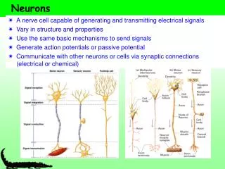
direction of information flow Postsynaptic Presynaptic neuron neuron little hill (base) (apex) Parts of two generalized CNS neurons Inhibitory terminal Excitatory terminal axon presynaptic terminal node of Ranvier initial myelin segment axon hillock cell body (soma) apical dendrites basal dendrites presynaptic terminal nucleus postsynaptic dendrite synaptic cleft Like Figure 2-1 (rotated)
Types of synapses • Synapses can form on dendrites, axons, soma, and even the presynapticteriminals • Two types of dendritic synapses are shown, one on spines, the other on the shaft • Type 1 synapses tend to be excitatory • Type 2 tend to be inhibitory Figure 10-3
Duration of postsynptic current all molecules begin here at t= 0 units: s-1 State 1 State 2 k21 open closed Concentration of acetylcholine at NMJ (because of acetylcholinesterase, turnover time ~ 100 μs) high 0 Number of open channels ms
At the nerve-muscle synapse, acetylcholinesterase is present at densities of > 1000 / μm2 near each synapse • This density is high enough to destroy each transmitter molecule as it leaves a receptor How are glutamate & GABA cleared from the synapse? • Na+ -coupled transporters for glutamate & GABA • are present at densities of > 1000 / μm2 near each synapse • This density is probably high enough to sequester each transmitter molecule as it leaves a receptor
Types of synaptic integration 1. Temporal 2. Spatial 3. Excitatory-inhibitory
Membrane capacitance integrates the current that goes across it This integration slows down voltage changes across the membrane Capacitive current is proportional to the derivative of voltage with respect to time Integrating both sides and solving for voltage we get, Figure 9-6
1B. Temporal Summation 2. Spatial summation Recording Recording Axon Axon Synaptic Current Synaptic Potential Long time constant (100 ms) Short time constant (20 ms) Synaptic Current Synaptic Potential Long length constant (1 mm) Short length constant (0.33 mm) ~ 100 pA Vm 2 mV 25 ms Vm Figure 10-14
1. Passive dendrites act like leaky cables Excitatory synapses V V EPSP measured in dendrite EPSP measured in soma Gulledge & Stuart (2005) J. Neurobiol 64:75,
Temporal and spatial integration of the EPSP from two synapses V V V Δt = 0 Δt = 0 Δt = 5 ms Prolonged rising phase Simultaneous, colocalized EPSPs (two individual trials) Nearly simultaneous, colocalized EPSPs (two individual trials) Simultaneous, Spatially distinct EPSPs Inspect the simulation, and run the movie, at http://www.neuron.yale.edu/neuron/static/about/stylmn.html Gulledge & Stuart (2005) J. Neurobiol 64:75,
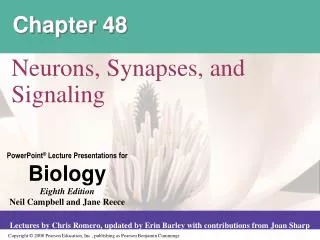
Active propagation in dendrites • Patch-clamp recordings from dendrites in brain slices show that dendrites can generate action potentials • A neuron was filled with a fluorescent dye • Voltage was recorded at two sites in current clamp mode, one in soma, one in dendrite • The occurrence of action potentials at the dendritic site shows that action potentials can actively propagate in dendrites Gulledge & Stuart (2005) J. Neurobiol 64:75,
Voltage-gated Na+channels in a dendrite 25 μm • Voltage-gated Na+ channels were recorded from a patch of membrane on the apical dendrite of a pyramidal neuron (left traces) • Step depolarization of the membrane patch opened the channels • The traces at the bottom left show the ensemble currents evoked by repeated voltage steps • Image in the middle shows the location of the recording site in a cell filled with fluorescent dye immunocytochemistry Averaged traces Now break the patch, to fill the cell with dye: Whitaker, Brain Res, 2001 Magee & Johnston, J Physiol (1995)
brain slice • Back propagation of the action potential • The existence of voltage-gated Na+ and Ca2+ channels in dendrites means that action potentials initiated at the axon hillock can “back propagate” to the dendrites • The back propagation of action potentials into the dendrites can have an important effect on synaptic signaling at excitatory synapses because NMDA receptors act as co-incidence detectors. Gulledge & Stuart (2005) J. Neurobiol 64:75,
Excitatory-inhibitory integration: • The “veto principle” of inhibitory transmission Inhibitory synapses work best when they are “near“ the excitatory event they will inhibit. “Near” means < one cable length. A. Inhibitory synapses on dendrites do a good job of inhibiting EPSPs on nearby spines B. Inhibitory synapses on cell bodies and initial segments do a good job of inhibiting spikes