Download
1 / 29
330 likes | 1.04k Views
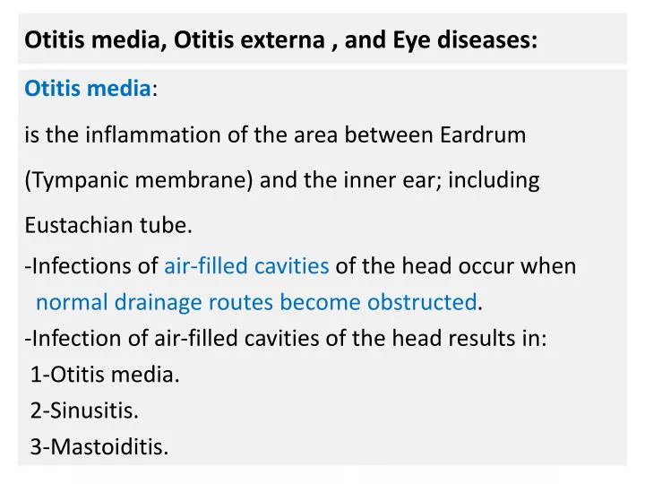
Otitis media, Otitis externa , and Eye diseases:. Otitis media : is the inflammation of the area between Eardrum (Tympanic membrane) and the inner ear; including Eustachian tube. -Infections of air-filled cavities of the head occur when normal drainage routes become obstructed .

E N D
Otitis media, Otitis externa , and Eye diseases: Otitis media: is the inflammation of the area between Eardrum (Tympanic membrane) and the inner ear; including Eustachian tube. -Infections of air-filled cavities of the head occur when normal drainage routes become obstructed. -Infection of air-filled cavities of the head results in: 1-Otitis media. 2-Sinusitis. 3-Mastoiditis.
N -Because the cavity of the middle ear is contiguous with the mastoid air cells(spaces of temporal bone); individuals with acute otitis media also have mastoiditis.
N -The majority of cases occur in children between 6 and 36 months of age. -Children are susceptible to otitis media for several reasons: 1-The medial orifice of the eustachian tube is more open in infancy than later in life. 2-Milkfeeding (giving a bottle at bedtime) results in reflux of pharyngeal contents into the lumen of eustachiantube. 3-Eustachiantube is shorter and more horizontal in young children. 4-The viral infection of upper respiratory tract and lymphoid tissue results in eustachian tube obstruction.
Pathogenesis: -Inflammation of upper respiratory tracts due to: 1-Viral infections; influenza A or B, and adenovirus. 2-Allergy(Rhinitis). -Swelling of lymphoid tissue (Eustachian tonsil)around eustachiantube. -Eustachian tube obstruction. -Absorption of air of middle ear slowly by surrounding tissues. -Creation of negative pressure (vacuum) in the middle ear. -Accumulation of fluids ; so normal flora of upper respiratory tract could invade middle ear space.
N -Colonization of middle ear cavity lining epithelium. -If the microbe has a polysaccharidecapsule: -Polyclonallymphocyteactivator; cytokines production; chemotaxisof immune cells and inflammation. -Conductive hearing loss. -If the infection is not treated; otitis media and mastoiditis could be complicated by: 1-Facialnerve paralysis. 2-Infection of peripheral nerves; results in deeperbrain abscess.
N 3-Infection of veins that bridge surrounding bony structures and the cerebral cortex; septic thrombophlebitis) results in subdural empyema (in some cases; related to epidural abscesses). Acute abscess is frequently caused by a mixed bacterial flora consisting of obligate and facultative anaerobic bacteria; similar to the mixture of microbes infecting middle ear, mastoid, and sinuses.
N Treatment of poly-microbial brain abscesses: Antibiotic combination: 1-Vancomycin or Ceftriaxone: to cover Staphylococci and other Gram positive beta-lactamase producers. 2-Metronidazole: to cover anaerobic bacteria. 3-Quinolones or Macrolides working effectively at acidic pH.
The Normal flora of upper respiratory tracts: -Streptococcus pneumoniae(Nasopharynx). -Haemophilusinfluenzae(non-type b) (Nasopharynx). -Moraxella catarrhalis(Nasopharynx). -Staphylococcus aureus. -Coagulase negative Staphylococcus species. -Diphtheroids species. -Neisseriae species. -Candida species.
Causes of Otitis media: 1-Streptococcuspneumoniae(the most common cause). 2-Non-typeable Haemophilusinfluenzae. (the second common cause). -Both Strep. pneumoniaeand H. influenzaecauses 80%of otitis media cases. 3-Moraxellacatarrhalis. -Gram’s negative non-motile coccobacilli in pairs. -Aerobic fastidious oxidase positive bacteria. 4-Other normal flora of upper respiratory tracts(rare).
Otitis Externa : Otitis Externa: is an inflammation of the outer ear and ear canal. -It is mainly caused by bacterial or fungal agents. -Otitis externa could be established due to: 1-Swimming in polluted water (germs contamination). 2-Impairment in the integrity of the skin (dermatitis). -Hospital acquired otitis externa could be caused by hospital dwelling bacteria as a post-surgical infection.
Causes of Otitis Externa: Exogenous: -Pseudomonas aeruginosa(the most common cause). Endogenous: Normal flora of outer ear canal: -Coagulase negative Staphylococci. -Staphylococcus aureus. -Gram negative bacilli. -Fungi: Candida species. Malassezia furfur.
N -Necrotizing otitis externais a form of infection in immunocompromised patients. -This type of infection could be complicated by inflammation of cranial nerves and their branches ; facial nerve paralysis. Diagnosis: Clinical specimen: Ear Swab (cotton swab).
N Culture: on Enriched media. -Blood agar incubated under aerobic conditions. -Chocolate agar: incubated under anaerobic conditions. Isolation of Pseudomonas species: -Encapsulated, motile, Gram negative bacilli. -Oxidase positive , Exopigments production. -Antibiotic resistance strains. (greenish yellowish exopigment production; pyoverdin).
N Isolation of Staphylococci: Staphylococcus aureus: Gram positive cocci, coagulase positive, novobiocin sensitive, and Mannitol fermenters. Other Staphylococci: Gram positive cocci, coagulase negative, novobiocin resistance. Novobiocin sensitivity Mannitol fermentation.
Infectious diseases of the Eye: Keratitis:inflammation of transparent eye’s cornea;the anterior part of the eye (covers the iris, and pupil). Causes: 1-Amoebic keratitis: a serious corneal infection usually affecting contact lens wearers. Etiology: Acanthamoeba. 2-Bacterial keratitis: Due to injury of wearing contact lenses. Etiology: Staphylococcus aureus, &Pseudomonas species.
N Staphylococcus aureus is a major cause of infections of the eyelid and cornea. Staphylococcus aureus can infect the glands of the eyelid; resulting in the production of a sty. Sty is a painful red swelling on the margin of the eyelid. Treatment: bacitracin ointment.
N 3-Fungal keratitis: Keratomycosis: Etiology: Fusariumspecies. -Infection is established due to corneal injury in agriculture workers or immunocompromised patients. FusariumChlamydospores. FusariumMacroconidia.
N 4-Viral Keratitis: Etiology: Herpes simplex virus types 1 and 2. Diseases: A-Primary infectious keratitis: Vesicular eruption of the eyelid, infection of cornea leading to corneal ulcers. B-Recurrent herpes keratitis: -(More common than primary keratitis). -In immunocompromised patients. -Mild irritation and photophobia.
N 5-Onchocercal Keratitis: Onchocerciasis: -Parasitic infection of the eye’s cornea (Corneal lesions). -Etiology: Onchocerca volvulus. -Transmitted by the bite of blackfly. -Disease: African River blindness.
Conjunctivitis: Conjunctiva: is a thin, translucent, mucous membrane that lines the eyelid and covers the white portion of the eyeball. Conjunctivitis is divided according to etiology into: A-Bacterial conjunctivitis: -Redness, swelling of the eyelid, and muco-purulent discharge. - Yellowish-greyish discharge: pyogenic cocci infection.
N Types of bacterial conjunctivitis: 1-Trachoma: Etiology: Chlamydiatrachomatis: Serotypes A, B, Ba, and C causes chronic keratoconjunctivitis (Trachoma) that results in blindness. Trachoma is a leading cause of blindness in endemic areas of northern India, the Middle East, and North Africa. Transmission: -Personal contact ; eye-to-eye via droplets by contaminated hands (transfer of elementary bodies).
N Chlamydiatrachomatis: -Unicellular obligatory intracellular bacteria that has rigid cell wall. -Infective stage: The elementary body. -Inclusion bodies (Trachoma) infected conjunctival epithelial cells (Reticulate body: diagnostic stage).
N 2-Ophthalmia neonatorum: Etiology: 1-Neisseriagonorrhoeae: -The most severe cause of hyperacute bacterial conjunctivitis of newborn. -It is acquired during passage of newborn through the birth canal of a mother infected by gonococci. -Neisseriaspecies are Gram negative oxidase positive diplococci that ferment glucose only.
N 2-Chlamydiatrachomatis:(Ophthalmianeonatorum): -This type of newborn conjunctivitis is associated with serotypes D-K. - 50% of Infants born with infection due to passage through the birth canal. - inclusion conjunctivitis heals without eye damage -Treatment of both types 1 and 2: Erythromycin ointment; most strains of Neisseria gonorrhoeae are Beta-Lactam resistant.
N B-Viral Conjunctivitis:(Pink Eye): diffuse pinkness of Conjunctiva. -Adenovirus infection is the most common cause of viral conjunctivitis. -Acute conjunctivitis, and pharyngoconjunctival fever. -A more serious infection is epidemic keratoconjunctivitis, which involves formation of a painful ulcer of the corneal epithelium. Electron microscopy: Double-Stranded DNA, Icosahedral naked virus.
N -Herpes simplex virus cause serious Herpetic keratoconjunctivitis, which requires treatment with acyclovir. -Acute hemorrhagic conjunctivitis is a highly contagious disease caused by: 1-Enterovirus 70. 2-Coxsackievirus A24. Viral hemorrhagic Conjunctivitis due to Enterovirus-70 infection.
Diagnosis of Eye infection: Clinical specimens: 1-Eye cotton Swab. 2-Conjunctival Scraping. 1-Direct microscopy: A-Swab for Microbiology (Gram’s stain ): detection of G+ve and G-ve bacteria, and yeast. B-Conjunctivalscrapes for cytology lab: detection of Chlamydiadiagnostic stage. C-ConjunctivalScrapes for immunohistochemistry: detection of viral infections or Chlamydia infection.
N 2-Detection of viral genetic material and Chlamydia genetic material by molecular methods: 1-Nucleic acid DNA hybridization.(Probe hybridization). 2-PCR : Primer amplification of genetic material. 3-Cultivation of bacterial agents: -Eye swab should be inoculated on Enriched media. -Blood agar and Chocolate agar should be incubated under aerobic and anaerobic conditions respectively. Clinical significance: isolation of pyogenic cocciand Neisseria gonorrhoeae.
Detection of Virus and Chlamydia genetic materials by Immunofluorescent Microscopy: Detection of viral antigen by Localization of specific Adenovirus- monoclonal antibodies. Coxsackievirus receptor on Epithelial cells of conjunctiva.