Download
1 / 55
550 likes | 616 Views
Lymphoproliferative diseases (44,9%) LEUKEMIAS (ALL, AML, CML, MDS) 33,6% LYMPHOMAS (HD, NHL) 9,8% HISTIOCYTOSIS 1,5% Solid tumours (55,1%) CNS TUMOURS 22,7%

E N D
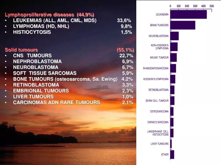
Lymphoproliferative diseases (44,9%) • LEUKEMIAS (ALL, AML, CML, MDS) 33,6% • LYMPHOMAS (HD, NHL) 9,8% • HISTIOCYTOSIS 1,5% Solid tumours (55,1%) • CNS TUMOURS 22,7% • NEPHROBLASTOMA 6,9% • NEUROBLASTOMA 6,7% • SOFT TISSUE SARCOMAS 5,9% • BONE TUMOURS (osteosarcoma, Sa. Ewing) 4,2% • RETINOBLASTOMA 3,3% • EMBRIONAL TUMOURS 2,3% • LIVER TUMOURS 1,0% • CARCINOMAS ADN RARE TUMOURS 2,1%
Treatment results of solid tumours in children • CNS TUMOURS 60% • NEPHROBLASTOMA 80% • NEUROBLASTOMA 45% • SOFT TISSUE SARCOMAS 43% • BONE TUMOURS 57% • RETINOBLASTOMA 70% • EMBRIONAL TUMOURS 85%
Brain tumors comprise approximately 20% of all childhood malignancies, second only to acute lymphoblastic leukemia in frequency.
Astrocytoma is the most common brain tumor, accounting for more than half of all primary central nervous system (CNS) malignancies.It can be distinguished: pilocytic astrocytoma(WHO grade I)diffuse astrocytoma(WHO grade II)anaplastic astrocytoma(WHO grade III)glioblastoma multiforme(WHO grade IV)
This MRI shows a juvenile pilocytic astrocytoma of the cerebellum This MRI shows a supratentorial glioblastoma multiforme
Medulloblastoma is the most common malignant brain tumor in children, accounting for 10-20% of primary central nervous system (CNS) neoplasms and approximately 40% of all posterior fossa tumors. It is a highly invasive embryonal neuroepithelial tumor that arises in the cerebellum and has a tendency to disseminate throughout the CNS early in its course. MRI showing a medulloblastoma of the cerebellum
Ependymoma is the third most common brain tumor in children, accounting for approximately 10% of primary central nervous system (CNS) neoplasms. It is a neuroepithelial tumor that arises within, or adjacent to, the ependymal lining of the ventricular system or the central canal of the spinal cord. It tends to invade locally, even if histological appearance is benign. Approximately 90% of tumors are intracranial, with up to 70% occurring in the posterior fossa. MRI showing an ependymoma of the fourth ventricle, compressing the cerebellum and brain stem
RETINOBLASTOMA Worldwide, the incidence of retinoblastoma is recorded to be about 11 cases per million children younger than 5 years. • Most cells comprising the tumor histologically resembled the cells of an undifferentiated retina of the embryo called retinoblasts. • The most widely held concept of histogenesis of retinoblastoma holds that it generally arises from a multipotential precursor cell (mutation in the long arm of chromosome 13 band 13q14) that could develop into almost any type of inner or outer retinal cell.
Retinoblastoma is diagnosed at an average of 18 months with 90% diagnosed before patients reach age 5 years. • Children who are affected bilaterally are diagnosed at an average age of 13 months, while patients with unilateral retinoblastoma are diagnosed at an average age of 24 months. • When a known family history of retinoblastoma exists, patients with bilateral retinoblastoma are diagnosed at an average age of 11 months.
Leukocoria (white pupillary reflex or cat's eye reflex) is the most common presenting sign, accounting for about 56.1% of cases. • Strabismus, which occurs as a result of visual loss, is the second most common mode of presentation. Thus, funduscopic examination through a well-dilated pupil must be performed in all cases of childhood strabismus. • Retinoblastoma can cause secondary changes in the eye, including glaucoma, retinal detachment, and inflammation secondary to tumor necrosis. • Pseudouveitis, with a red eye and pain associated hypopyon and hyphema, is a rare presentation. It is characteristic of an infiltrating type of retinoblastoma in which the tumor cells invade the retina diffusely without forming a discrete tumor mass. • Orbital inflammation mimicking orbital cellulitis may occur in eyes with necrotic tumors and does not necessarily imply extraocular extension. • Proptosis is a more common presenting symptom in most underdeveloped countries.
EBRT is still indicated in selected circumstances: For eyes with significant vitreous seeding For children who have progression of disease while undergoing chemoreduction For tumors extending up to or beyond the cut margin of the optic nerve of an enucleated eye Radioactive isotope plaques Use of either radioactive 60 Co (cobalt), radioactive 125 I (iodine), which is presently the most used, radioactive 192 Ir (iridium), and radioactive 106 Ru (ruthenium) Radioactive 125 I plaque treatment is recommended for treatment of one larger tumor or a limited number of moderately sized tumors (<3) present in noncritical areas Advantage - Locally directed treatment to the tumor minimizing radiation to the normal tissue Disadvantage - Incomplete treatment, high dose to local sclera, significantly less irradiation for anterior lesions, and difficulty placing posterior plaques
Enucleation • Enucleation is performed when there is no chance of preserving useful vision in an eye. • Patients generally requiring enucleation are those who present with total retinal detachments and/or the posterior segment is full of the tumor in which case it is clear the patient cannot retain any form of useful vision. • Cryotherapy • Cryotherapy can be used primarily for small anteriorly located tumors, remote from the disc and macula but also may be indicated for recurrence after radiation therapy. • Cryotherapy is performed transsclerally. Under direct visualization, freezing is carried out until the ice ball incorporates the entire tumor. A refreeze-thaw cycle is repeated 3-4 times. • Complete disappearance of the tumor with a flat pigmented scar is the sign of successful treatment. This can be repeated if the tumor does not respond initially. • Photocoagulation • Photocoagulation can be used as primary therapy for small posteriorly located tumors. • There is a danger of producing large field defects near the disc and decreased vision resulting from macular pucker by photocoagulation near the macula. • The technique is performed by placing a double row of confluent burns around each tumor using a photocoagulator. • It is important not to do direct treatment on the tumor itself because the light color of the tumor generally precludes absorption of sufficient energy and there is a danger of exploding the tumor with spread of viable tumor debris into the vitreous and other parts of the retina. • Successful treatment with photocoagulation takes weeks to evolve, which is a complete disappearance of the tumor and replaced with a flat area. • Photocoagulation also can be used for tumor recurrences after EBRT.
Objawy kliniczne • Endophytic growth occurs when the tumor breaks through the internal limiting membrane and has an ophthalmic appearance of a white-to-cream mass showing either no surface vessels or small irregular tumor vessels. This growth pattern typically is associated with vitreous seeding wherein small fragments of tissue become separated from the main tumor. • Exophytic growth occurs in the subretinal space. This growth pattern often is associated with subretinal fluid accumulation and retinal detachment. • Diffuse infiltrating growth is a rare subtype comprising 1.5% of all retinoblastoma. It is characterized by a relatively flat infiltration of the retina by tumor cells but without a discrete tumor mass.
Nephroblastoma • third most common solid tumour (6-9 cases/ 1 million children) • location • in one kidney (90-95%) • in both kidneys (5-10%) • hereditary tumours (7-10%) • WT1 gene (11p13), WT2 gene (11p15), WT3 gene (?)
The most common presentation of Wilms tumor is the presence of an asymptomatic abdominal mass. • Hypertension, gross hematuria, and fever are observed in 5-30% of patients. • A small number of patients who have hemorrhaged into their tumor may present with signs of hypotension, anemia, and fever. • Rarely, patients with advanced-stage disease may present with respiratory symptoms related to the presence of lung metastases.
CT scan of a patient with a right sided favorable histology Wilms tumor • CT scan of child with Stage IV favorable histology Wilms tumor. Note the presence of bilateral pulmonary metastases
Nephroblastoma • clinical staging : SIOP system and NWTS (stages I-V) • histological classification according to Beckwith and Palmer • blastomatous element • epithelial (tubular) elements • mesenchymal elements
-large tongue (macroglossia)-large organs (visceromegaly)-large body size (macrosomia)-hernia of the navel-small head (microcephaly)-Wilms’ tumour • Cytogenetics studies: • An 11p13 deletion as in the WAGR syndrome (Wilms, aniridia, genitourinary abnormalities, mental retardation) • A duplication of the paternal allele 11p15 as in BWS • Mutational analysis of the WT1 gene in cases where Denys-Drash syndrome (intersexual disorders, nephropathy, Wilms tumor) is suspected
Neuroblastoma • second most common solid tumour (1 case/ 10000 children) • location • retroperineum (75-80%) • posterior mediastinum (20%) • cervical ganglia (5%) • clinical appearance depends on the origin of the tumor, metastasis and hormonally active metabolic products
Age, stage, and some molecular defects encountered in tumor cells are important prognostic factors and are used for risk-stratification and treatment assignment. • The differences in outcome for patients with neuroblastoma are striking. Infants younger than 1 year have a good prognosis, even in the presence of metastatic disease, whereas older patients with metastatic disease fare poorly, even when treated with aggressive therapy. • Unfortunately, approximately 70-80% of patients older than 1 year present with metastatic disease, usually to lymph nodes, liver, bone, and bone marrow.
More than 90% of patients have elevated homovanillic acid (HVA) and/or vanillylmandelic acid (VMA) detectable in urine. • Mass screening studies using urinary catecholamines in neonates and infants in Japan, Quebec, and Europe have demonstrated the ability to detect neuroblastoma before it clinically is apparent. However, most of the tumors identified using this method occur in infants who have a good prognosis. No data exist to suggest that mass screening has decreased deaths from high-risk neuroblastoma. • Markers associated with a poor prognosis are elevated ferritin, serum lactate dehydrogenase (LDH), and serum neuron-specific enolase (NSE).
MRI of a left adrenal mass. The mass was detected by fetal ultrasound at 30 weeks of gestation. During infancy, the mass was found on the inferior pole of the left adrenal and was completely resected. Before surgery, the metastatic workup was negative. Surgical pathology service confirmed a diagnosis of neuroblastoma. After 3 years of follow-up care, no recurrence was observed.
A one-week-old neonate had an abdominal ultrasound for evaluation of projectile vomiting. A right adrenal mass (100% cystic) was an incidental finding. Evaluation of the mass by CT was consistent with an adrenal bleed (3.6 x 3.1 x 2.4 cc). The infant was followed at 2 weeks (2D size diminished to 1.5 x. 2.4 cm sq. by Ultrasound) and then at 6 weeks to document that the adrenal bleed continued to involute. Urine catecholamines were normal.
Symptoms include abdominal pain, emesis, weight loss, anorexia, fatigue, bone pain, and chronic diarrhea. Hypertension is an uncommon sign of the disease and generally is caused by renal artery compression, not catecholamine excess. • Because more than 50% of patients present with advanced-stage disease, usually to the bone and bone marrow, the most common presentation includes bone pain and a limp. However, patients also may present with unexplained fever, weight loss, irritability, and periorbital ecchymosis secondary to metastatic disease to the orbits. The presence of bone metastases can lead to pathologic fractures. • Approximately two thirds of patients with neuroblastoma have abdominal primaries. In these circumstances, patients can present with an asymptomatic abdominal mass that usually is discovered by the parents or a caregiver. • Symptoms produced by the presence of the mass depend on its proximity to vital structures and usually progress over time. • Tumors arising from the paraspinal sympathetic ganglia can grow through the spinal foramina into the spinal canal and impinge on the spinal cord. This may result in the presence of neurologic symptoms, including weakness, limping, paralysis, and even bladder and bowel dysfunction. • Thoracic neuroblastomas (posterior mediastinum) may be asymptomatic and usually are diagnosed by imaging studies obtained for other reasons. Presenting signs or symptoms may be insignificant and involve mild airway obstruction or chronic cough, leading to a chest radiograph. • Thoracic tumors extending to the neck can produce Horner syndrome. Primary cervical neuroblastoma is rare but should be considered in the differential diagnosis of masses of the neck, especially in infants younger than 1 year with feeding or respiratory difficulties.
Neuroblastoma • clinical staging : Evans’ system and INNS (International Neuroblastoma Saging system) • histological staging according to Hughes’ or Shimada’s classification
N-MYC, RAS amplification and expression • 1p deletion • 17q • 11q, 14q deletion • Exspression of TRKA, TRKB, TRKC (receptor for neurotropins)
Liver tumours • Hepatoblastoma • usually occurs in children under 5 years of age • hepatocellular carcinoma • usually occurs in older children • the cause of cancer : chronic liver disease, viral hepatitis, hemochromatosis, mycotoxins • AFP serum level - indicator • whether the disease is responding to treatment • whether the disease is come back
Hepatoblastoma usually is diagnosed as an asymptomatic abdominal mass. Approximately 10% of patients have incidental findings of hemihypertrophy. Hepatoblastoma can be associated with isosexual precocity. Penile and testicular enlargement without pubic hair is seen in patients with tumors that secrete the b subunit of human chorionic gonadotropin (b-hCG). HCC usually presents as a slowly enlarging, right, upper-quadrant mass that may be found during a routine physical examination, brought to medical attention by the patient, or discovered by the patient's parents. Many children also experience localized pain, nausea, vomiting, and weight loss. Nearly 25% of patients present with jaundice. In adults, chronic hepatitis secondary to alcohol exposure, infection with hepatitis, and hereditary hemochromatosis are predisposing factors. Aflatoxins and other environmental factors also are likely to play a role in the pathogenesis in adults. In comparison, children are far more likely to have inherited errors of metabolism, such as tyrosinemia or urea cycle enzymopathies. Liver diseases that cause cirrhosis increase risk for developing HCC. Children with biliary atresia, chronic cholestasis, or glycogen storage diseases are at increased risk. Symptoms can be masked in children with preexisting hepatic diseases, and, accordingly, a change in a chronic disease pattern merits careful consideration for the possibility of a new malignancy.
Computed tomogram of hepatoblastoma Computed tomogram of large hepatocellular carcinoma
Osteosarcoma • usually associated with the period of rapid growth in adolescence • most common malignant bone tumour in children • Rb gene is associated with osteosarcoma • tumour tends to occure in the femur (distal end), humerus (proximal end) and tibia (proximal end) • can occur in any bone
Lateral plain radiograph of the knee shows an osteosarcoma of the distal femur. The lesion is mainly posterior, with disruption and elevation of the periosteum (Codman triangle), and extends beyond the bone into the soft tissue. MRI of the same distal femoral osteosarcoma
Ewing’s sarcoma • usually associated with the period of rapid growth in adolescence • tumour tends to occure in the long bones of the extremities, the pelvis or the chest • may occur in the skull or flat bones of the trunk • can occur in any bone • the tumour spreads easily, often to lungs and other bones (metastases present in 1/3 of the children at the time of diagnosis)
Rotationplasty in th epatient with osteosarcoma of the distal femur
1. TERATOMA • 2. GERMINOMA (dysgerminoma and seminoma) • 3. CARCINOMA EMBRYONALE • 4. YOLK SAC TUMOR • 5. CHORIOCARCINOMA • 6. POLYEMBRYOMA • 7. GONADOBLASTOMA
Germ cell tumors account for 95% of testicular tumors and include seminomas, teratomas, choriocarcinomas, and mixed tumors. • Seminomas comprise approximately 50% of all germ cell tumors. Seminomas are generally believed to arise from the germinal epithelium of the seminiferous tubules because "seminoma cells" are morphologically similar to spermatogonia and also because seminomas are frequently found within the seminiferous tubules in early stages.