Download
1 / 31
310 likes | 315 Views
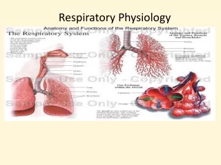
BI 233 Laboratory Respiratory Physiology. Respiratory Terminology. Pulmonary ventilation Process by which O₂ enters and CO₂ exits alveoli (breathing). Respiration Process by which O₂ and CO₂ diffuse in and out of the blood (gas exchange) Occurs in 2 areas of the body.

E N D
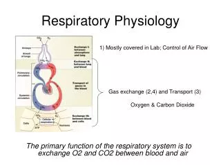
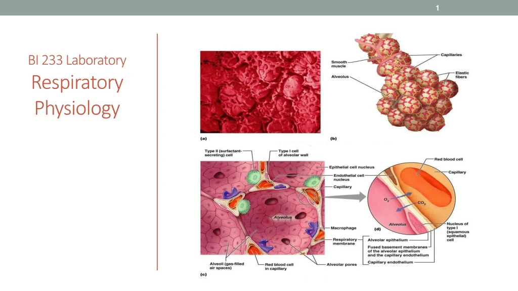
Respiratory Terminology • Pulmonary ventilation • Process by which O₂ enters and CO₂ exits alveoli (breathing). • Respiration • Process by which O₂ and CO₂ diffuse in and out of the blood (gas exchange) • Occurs in 2 areas of the body. • External respiration – gas exchange across respiratory membrane of lungs (alveoli) into the blood. • Internal respiration – gas exchange between the blood and the interstitial fluid, tissues and cells • Cellular respiration • Series of metabolic processes where living cells produce energy through oxidation of organic substances.
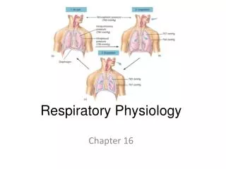
Pulmonary Ventilation:Respiratory Cycle • Ventilation is breathing with a respiratory cycle including:. • Inspiration • Air moves into lungs when pressure inside lungs is less than atmospheric pressure. • Expiration • Air moves out of the lungs when pressure inside lungs is greater than atmospheric pressure. • 1 ATM or 760mm Hg
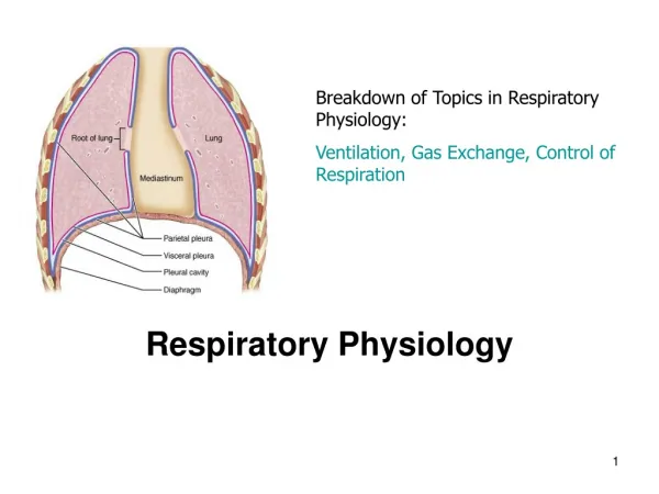
Pneumothorax • Pleural cavities are sealed cavities not open to the outside. • Presence of air in pleural cavity. • Loss of negative intrapleural pressure allows lungs to recoil and collapse. • Collapse of lung (or part of lung) is called atelectasis. • Injuries to the chest wall that let air enter the intrapleural space. • Mechanism of injury. • Collapsed lung on same side as injury. • Surface tension and recoil of elastic fibers causes the lung to collapse.
Spirometry • Is a common test used to assess how well your lungs work by measuring how much air you inhale, how much you exhale and how quickly you exhale. • It is used to diagnose asthma, chronic obstructive pulmonary disease (COPD) and other conditions that affect breathing
Pulmonary Volumes (how much) • Exchange of O₂ and CO₂ dependent on normal volumes of air moving in and out, and remaining volumes. • Respiratory volumes • Tidal volume (TV) • Air exhaled after a normal inspiration (quiet breathing). • Inspiratory reserve volume (IRV) • Air in excess of tidal inspiration that can be inhaled with maximum effort. • Normal = 3.3L • Expiratory reserve volume (ERV) • Air in excess of tidal expiration that can be exhaled with maximum effort. • Normal = between 1.0 and 1.2L • Residual volume (RV) • Air remaining in lungs after maximum expiration, keeps alveoli inflated. • Normal = 1.1-1.2L
Pulmonary Capacities • Vital capacity (VC) • Amount of air that an be exhaled with maximum effort after maximum inspiration. • Influenced by size of thoracic cavity and posture. • Assess strength of thoracic muscles and pulmonary function. • VC = IRV + TV + ERV • Inspiratory capacity (IC) • Maximum amount of air that can be inhaled after a normal (tidal) expiration. • Functional residual capacity (FRC) • Amount of air in lungs after a normal respiration (tidal). • Total lung capacity (TLC) • Maximum amount of air lungs can hold. • Sum of all four lung volumes. • TLC = TV + IRV + ERV + RV
Pulmonary Volumes, Capacities and Air Flow • Wet Spirometers -measure volumes of gas that lungs exhale, can obtain ERV, TV and VC function of time. Can calculate the IRV using the following formula: IRV= VC-(ERV+TV)
Affects on Respiratory Volumes and Capacities • Age • lung compliance, respiratory muscles weaken. • Exercise • Maintains strength of respiratory muscles. • Body size • Proportional, big body has large lungs. • Restrictive disorders • compliance and vital capacity (volume). • Obstructive disorders • Interfere with airflow, expiration requires more effort or less complete.
FEV: Most Important Measurement of Lung Function • Forced expiratory volume (FEV) – volume of gas expired over time – (typically 1 sec=FEV1) • AKA: forced vital capacity (FVC) • Maximum volume of gas expired/second during forced expiration (% of VC). • Exhaled from full inhalation as forcefully and rapidly as possible. • Amount of air exhaled may be measured during first (FEV1), second (FEV2), and/or third seconds (FEV3) of forced breath. • Healthy adult - 75 to 85% in 1 sec. • FEV used to… • Diagnose obstructive lung diseases (asthma and COPD). • Individuals with asthma or COPD have lower FEV1 result than a healthy person. • Indicate effectiveness of medications prescribed for lung disease. • Monitor disease status. • Decreases in FEV1 value may indicate condition is worsening.
Respiratory Diseases • Obstructive Lung Disease – (Outflow) • Not getting air out • Large over inflated lungs because air can’t get out • Asthma (airways spasm and close) • Chronic bronchitis • Emphysema (lungs loose their elasticity) • Restrictive Lung Disease – (Intake) • No problem expiring air there is just less air • Lungs can not expand (an intake problem) • Fibrosis (tissue gets stiff) • Muscular diseases (muscular dystrophy, ALS) • obesity
FEV1Forced Expiratory Volume = volume of air exhaled under forced conditions in the first one second FEV1 /VC Percent Forced Vital Capacity = the ratio of FEV1 to VC. In healthy adults this should be approximately 70–85% (declining with age). In obstructive lung disease, (asthma, COPD, chronic bronchitis, emphysema) the FEV1 is reduced because of increased airway resistance to expiratory flow. Thus, the FEV1/FVC ratio will be reduced. In restrictive lung disease, (fibrosis) the FEV1 and VC are equally reduced. Thus, the FEV1/VC ratio should be approximately normal.
Activities • Spirometry exercise – pg. 203-205
What happens if we can’t expire CO2? • Hypercapnia (too much CO2) • If CO2 accumulates in the bloodstream the blood becomes acidic a condition called respiratory acidosis. • Hypocapnia (too little CO2) • If there is not enough CO2 in the bloodstream then blood becomes alkaline a condition call respiratory alkalosis.
Gas Exchange and Transport Lungs decrease acid y incresing respiration or increase acid by decreasing respiration Kidneys decrease acid by urinating H+ and putting base into blood
Gas Exchange and Transport: CO₂ • CO₂ is carried in blood 3 ways: • Small percentage CO2 dissolves into plasma (7%). • Hemoglobin can bind to some CO2(23%). • Most interacts with plasma H2O to convert to carbonic acid (H2CO3) (70%). • Brain translates an increase in CO2as not breathing enough. • Carbon dioxide in tissues. • PCO₂ = 45 mmHg • Enters blood and combines with water to form carbonic acid. • Carbonic acid splits into hydrogen ion (H⁺) and bicarbonate (HCO3¯). • HCO3¯ + H⁺ forms H2CO3 • H2CO3splits to form H2O and CO2 • CO2 diffuses from blood into alveoli.
Effects of Hydrogen Ions • Respiratory alkalosis (pH > 7.45) • Hypocapnia (PCO2) < 37 mmHg. • Caused by hyperventilation • Corrected by hypoventilation, pushes reaction to the right. CO2 + H2O H2CO3 HCO3-+ H+ • H+, lowers pH to normal. • Respiratory acidosis (pH < 7.35) • Caused by failure of pulmonary ventilation - hypoventilation. • Hypercapnia (PCO2) > 43 mmHg • CO2 easily crosses blood-brain barrier, in CSF the CO2 reacts with water and releases H+, central chemoreceptors strongly stimulate inspiratory center. • Corrected by hyperventilation, pushes reaction to the left by “blowing off ” CO2 (expired). CO2+ H2O H2CO3 HCO3-+ H+
Buffers • Any mechanism that resists changes in pH by converting a strong acid or base to a weak one. • A chemical buffer is a substance that binds H+ and removes it from solution as its concentration begins to rise, or releases H+ into solution as its concentration falls
Gas Exchange and Transport Lungs decrease acid by increasing respiration or increase acid by decreasing respiration Kidneys decrease acid by urinating H+ and putting base into blood
Do Buffer Activity in lab manual • Follow the instructions in your manual
Activity: Effect of pCO2 on respiratory Rate • 1. Observe lab partner for one minute and record their respiratory rate (# of breaths per min) • 2. Next have partner take slow, deep breaths for two minutes and record respiratory rate for one minute • Was there an increase or decrease in the respiratory rate? • 3. Next have partner breathe in and out of a paper bag (covering their nose and mouth with the bag) for two minutes and then record respiratory rate for one minute. • Was there an increase or decrease in the respiratory rate?
Activity: Effect of CO2 on the pH of a solution • Since we can’t measure the effects of ventilatory rate on blood pH directly in the lab because it requires an analysis of arterial blood we will use water and test the pH while resting, exercising and after exercising. • Follow the instructions in your lab manual and then answer the questions.
Oxygen Imbalances: Hypoxia • Hypoxemic hypoxia - usually due to inadequate pulmonary gas exchange • High altitudes, drowning, aspiration, respiratory arrest, degenerative lung diseases, CO poisoning. • Ischemic hypoxia - inadequate circulation. Shock • Anemic hypoxia - anemia • Histotoxic hypoxia - metabolic poison (cyanide). • Cyanosis- blueness of skin. • Primary effect of hypoxia is tissue necrosis, organs with high metabolic demands affected first.
Activity: Pulse Oximeter • Check your oxygen saturation levels and record on chart
Works Cited Marieb, E. N. (2012). Essentials of human anatomy & physiology (6th ed.). Boston: Pearson Education, Inc. Marieb, E.N., Mitchell, S.J. & Smith, L.A. (2012). Human anatomy and physiology laboratory manual (10th ed.). Boston: Pearson Education, Inc. Martini, F., Nath, J. & Bartholomew, E.F. (2012). Fundamentals of anatomy & physiology (9th ed.). Boston: Pearson Education, Inc. McPhee, J. & Papadakis, M. (2012) Current medical diagnosis & treatment (51st ed.). New York: McGraw Hill. Patton, T. & Thibodeau, G. (2013). Anatomy & physiology (8th ed.). St. Louis:Mosby Elsevier. Saladin, K. S. (2012). Anatomy & physiology: The unity of form and function (6th ed.). New York: McGraw Hill. Tortora, G.J. & Derrickson, B.H. (2012). Principles of anatomy and physiology (13th ed.). Hoboken, NJ: Wiley