Download
1 / 49
490 likes | 496 Views
МАССПЕКТРОМЕТРИЯ. ФИЗИКОХИМИЧЕН МЕТОД ЗА ИЗСЛЕДВАНЕ НА ВЕЩЕСТВАТА ПОСРЕДСТВОМ ЙОНИЗИРАНЕТО ИМ И ПОСЛЕДВАЩОТО РАЗДЕЛЯНЕ НА ПОЛУЧЕНИТЕ ЙОНИ В ЗАВИСИМОСТ ОТ ВЕЛИЧИНАТА НА ТЯХНАТА МАСА.

E N D
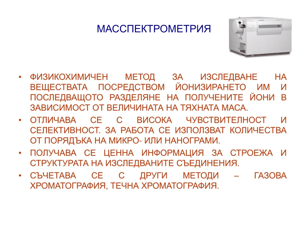
МАССПЕКТРОМЕТРИЯ • ФИЗИКОХИМИЧЕН МЕТОД ЗА ИЗСЛЕДВАНЕ НА ВЕЩЕСТВАТА ПОСРЕДСТВОМ ЙОНИЗИРАНЕТО ИМ И ПОСЛЕДВАЩОТО РАЗДЕЛЯНЕ НА ПОЛУЧЕНИТЕ ЙОНИ В ЗАВИСИМОСТ ОТ ВЕЛИЧИНАТА НА ТЯХНАТА МАСА. • ОТЛИЧАВА СЕ С ВИСОКА ЧУВСТВИТЕЛНОСТ И СЕЛЕКТИВНОСТ. ЗА РАБОТА СЕ ИЗПОЛЗВАТ КОЛИЧЕСТВА ОТ ПОРЯДЪКА НА МИКРО- ИЛИ НАНОГРАМИ. • ПОЛУЧАВА СЕ ЦЕННА ИНФОРМАЦИЯ ЗА СТРОЕЖА И СТРУКТУРАТА НА ИЗСЛЕДВАНИТЕ СЪЕДИНЕНИЯ. • СЪЧЕТАВА СЕ С ДРУГИ МЕТОДИ – ГАЗОВА ХРОМАТОГРАФИЯ, ТЕЧНА ХРОМАТОГРАФИЯ.
МАССПЕКТРОМЕТРИЯ • ПРИНЦИПНА ПРОЦЕДУРА: • ВЪВЕЖДАНЕ НА ВЕЩЕСТВОТО; • ЙОНИЗАЦИЯ НА НЕУТРАЛНИТЕ МОЛЕКУЛИ; • РАЗПРЕДЕЛЕНИЕ НА ЙОНИТЕ В ЗАВИСИМОСТ ОТ МАСАТА ИМ; • РЕГИСТРАЦИЯ НА ЙОНИТЕ.
(Ph. Eur. method 2.2.43) Mass spectrometry is based on the direct measurement of the ratio of the mass to the number of positive or negative elementary charges of ions (m/z) in the gas phase obtained from the substance to be analysed. This ratio is expressed in atomic mass units (1 a.m.u. = one twelfth the mass of 12C) or in daltons (1 Da = the mass of the hydrogen atom). The ions, produced in the ion source of the apparatus, are accelerated and then separated by the analyser before reaching the detector. All of these operations take place in a chamber where a pumping system maintains a vacuum of 10-3 to 10-6 Pa. The resulting spectrum shows the relative abundance of the various ionic species present as a function of m/z. The signal corresponding to an ion will be represented by several peaks corresponding to the statistical distribution of the various isotopes of that ion. This pattern is called the isotopic profile and (at least for small molecules) the peak representing the most abundant isotopes for each atom is called the monoisotopic peak. Information obtained in mass spectrometry is essentially qualitative (determination of the molecular mass, information on the structure from the fragments observed) or quantitative (using internal or external standards) with limits of detection ranging from the picomole to the femtomole.
МАССПЕКТРОМЕТРИЯ • ЙОНИЗИРАНЕ НА МОЛЕКУЛИТЕ: - ЙОНИЗИРАНЕ ЧРЕЗ ЕЛЕКТРОНЕН УДАР – ОТДЕЛНАТА МОЛЕКУЛА ВЗАИМОДЕЙСТВА С ЕЛЕКТРОН С ПО-ВИСОКА ЕНЕРГИЯ, КОЙТО ВЪЗБУЖДА ЕЛЕКТРОННАТА ОБВИВКА НА МОЛЕКУЛАТА. ОТ НЕЯ СЕ ОТДЕЛЯ ЕДИН ЕЛЕКТРОН И СЕ ПОЛУЧАВА ПОЛОЖИТЕЛНО ЗАРЕДЕН ЙОН. - ЕНЕРГИЯТА, НЕОБХОДИМА ЗА ОТДЕЛЯНЕ НА ЕЛЕКТРОН ОТ НАЙ-ВЪНШНАТА МОЛЕКУЛНА ОРБИТА, ЗА ПО-ГОЛЯМА ЧАСТ ОТ ОРГАНИЧНИТЕ СЪЕДИНЕНИЯ Е ОТ 7 ДО 13 еV. ПРИ ЕНЕРГИЯ НА ЕЛЕКТРОНИТЕ ПО-МАЛКА ОТ ЙОНИЗАЦИОННИЯ ПОТЕНЦИАЛ НА МОЛЕКУЛАТА, НЕ НАСТЪПВАТ ПРОМЕНИ.
Потокът от електрони генерира йоните М+ в разнообразни енергетични състояния. Някои йони се получават с много голяма вътрешна енергия (ротационна, вибрационна, електронна), която се изразходва при реакциите на фрагментиране и се получават йони с по-малки маси. Други йони се съпротивляват на разграждането, тъй като са образувани с недостатъчно енергия за фрагментиране. Повечето процеси на фрагментация са ендотермични и поради това йоните с ниска енергия не се разпадат в източника и се детектират като непроменени молекулни маси.
МАССПЕКТРОМЕТРИЯ - ПРИ ОБРАЗУВАНЕ НА МОЛЕКУЛЕН ЙОН, ЙОНИЗАЦИЯТА ПРОТИЧА БЕЗ РАЗКЪСВАНЕ НА МОЛЕКУЛАТА: M + e- = M+ + 2e- ПРИНЦИПНО Е ВЪЗМОЖНО И ПОЛУЧАВАНЕТО НА ОТРИЦАТЕЛЕН ЙОН ПРИ ПРОЦЕС НА ЗАХВАЩАНЕ НА ЕЛЕКТРОН ОТ МОЛЕКУЛАТА. ПРИ ЕДНАКВИ УСЛОВИЯ ПОЛУЧАВАНЕТО НА ОТРИЦАТЕЛНИ ЙОНИ Е МНОГОКРАТНО ПО-МАЛКО. ПРИ ПОВИШАВАНЕ НА ЕНЕРГИЯТА НА ЕЛЕКТРОННИЯ СНОП, НАРАСТВА ВЕРОЯТНОСТТА ЗА ЙОНИЗАЦИЯ. КОГАТО СВОБОДНАТА ЕНЕРГИЯ НА МОЛЕКУЛНИЯТ ЙОН ДОСТИГНЕ СТОЙНОСТТА НА СЪОТВЕТНАТА ДИСОЦИАЦИОННА ЕНЕРГИЯ, НАСТЪПВА РАЗКЪСВАНЕ НА МОЛЕКУЛАТА И ОБРАЗУВАНЕ НА ЙОННИ ФРАГМЕНТИ.
МАССПЕКТРОМЕТРИЯ - НАЧИНЪТ НА РАЗПАДАНЕТО НА МОЛЕКУЛНИЯ ЙОН ЗАВИСИ ОТ СТРУКТУРАТА НА МОЛЕКУЛАТА, ОТ ВИДА НА ФУНКЦИОНАЛНИТЕ ГРУПИ И ТЯХНОТО РАЗПОЛОЖЕНИЕ. - ОСВЕН ЧРЕЗ ЕЛЕКТРОНЕН УДАР СЕ ИЗПОЛЗВАТ И ДРУГИ МЕТОДИ КАТО ФОТОЙОНИЗАЦИЯ, ХИМИЧНА И ПОЛЕВА ЙОНИЗАЦИЯ, ЙОНИЗАЦИЯ С БЪРЗИ АТОМИ, ЛАЗЕРНА ЙОНИЗАЦИЯ. - ХИМИЧНАТА ЙОНИЗАЦИЯ СЕ ОСЪЩЕСТВЯВА ПРИ ВЗАИМОДЕЙСТВИЕ НА МОЛЕКУЛИТЕ НА ВЕЩЕСТВОТО С ЙОНИТЕ НА ПРЕДВАРИТЕЛНО ЙОНИЗИРАН ГАЗ (ГАЗ-РЕАГЕНТ). ПРИ GC/MS РОЛЯТА НА ГАЗ-РЕАГЕНТ МОЖЕ ДА СЕ ИГРАЕ ОТ ГАЗА-НОСИТЕЛ.
Modes of ionisation Electron impact The sample, in the gas state, is ionised by a beam of electrons whose energy (usually 70 eV) is greater than the ionisation energy of the sample. In addition to the molecular ion M+, fragments characteristic of the molecular structure are observed. This technique is limited mainly by the need to vaporise the sample. This makes it unsuited to polar, heat-labile or high molecular mass compounds. Electron impact is compatible with the coupling of gas chromatography to mass spectrometry and sometimes with the use of liquid chromatography. Chemical ionisation This type of ionisation involves a reagent gas such as methane, ammonia, nitrogen oxide, nitrogen dioxide or oxygen. The spectrum is characterised by ions of the (M + H)+ or (M - H)- types, or adduct ions formed from the analyte and the gas used. Fewer fragments are produced than with electron impact. A variant of this technique is used when the substance is heat-labile: the sample, applied to a filament, is very rapidly vaporised by the Joule-Thomson effect (desorption chemical ionisation).
Fast-atom bombardment (FAB) or fast-ion bombardment ionisation (liquid secondary-ion mass spectrometry LSIMS) The sample, dissolved in a viscous matrix such as glycerol, is applied to a metal surface and ionised by a beam of neutral atoms such as argon or xenon or high-kinetic-energy caesium ions. Ions of the (M + H)+ or (M - H)– types or adduct ions formed from the matrix or the sample are produced. This type of ionisation, well suited to polar and heat-labile compounds, allows molecular masses of up to 10 000 Da to be obtained. The technique can be combined with liquid chromatography by adding 1 per cent to 2 per cent of glycerol to the mobile phase; however, the flow rates must be very low (a few microlitres per minute). These ionisation techniques also allow thin-layer chromatography plates to be analysed by applying a thin layer of matrix to the surface of these plates. Field desorption and field ionisation The sample is vaporised near a tungsten filament covered with microneedles (field ionisation) or applied to this filament (field desorption). A voltage of about 10 kV, applied between this filament and a counter-electrode, ionises the sample. These two techniques mainly produce molecular ions M+, and (M + H)+ ions and are used for low polarity and/or heat-labile compounds.
Matrix-assisted laser desorption ionisation (MALDI) The sample, in a suitable matrix and deposited on a metal support, is ionised by a pulsed laser beam whose wavelength may range from UV to IR (impulses lasting from a picosecond to a few nanoseconds). This mode of ionisation plays an essential role in the analysis of very high molecular mass compounds (more than 100 000 Da) but is limited to time-of flight analysers (see below). Electrospray This mode of ionisation is carried out at atmospheric pressure. The samples, in solution, are introduced into the source through a capillary tube, the end of which has a potential of the order of 5 kV. A gas can be used to facilitate nebulisation. Desolvation of the resulting microdroplets produces singly or multiply charged ions in the gas phase. The flow rates vary from a few microlitres per minute to 1 ml/min. This technique is suited to polar compounds and to the investigation of biomolecules with molecular masses of up to 100 000 Da. It can be coupled to liquid chromatography or capillary electrophoresis.
Atmospheric-pressure chemical ionisation (APCI) Ionisation is carried out at atmospheric pressure by the action of an electrode maintained at a potential of several kilovolts and placed in the path of the mobile phase, which is nebulised both by thermal effects and by the use of a stream of nitrogen. The resulting ions carry a single charge and are of the (M + H)+ type in the positive mode and of the (M - H)– type in the negative mode. The high flow rates that can be used with this mode of ionisation (up to 2 ml/min) make this an ideal technique for coupling to liquid chromatography. The sample, in the mobile phase consisting of water and organic modifiers and containing a volatile electrolyte (generally ammonium acetate) is introduced in nebulised form after having passed through a metal capillary tube at controlled temperature. Acceptable flow rates are of the order of 1 ml/min to 2 ml/min. The ions of the electrolyte ionise the compounds to be analysed. This ionisation process may be replaced or enhanced by an electrical discharge of about 800 volts, notably when the solvents are entirely organic. This technique is compatible with the use of liquid chromatography coupled with mass spectrometry.
Alkanes: Simple alkanes tend to undergo fragmentation by the initial loss of a methyl group to form a (m-15) species. This carbocation can then undergo stepwise cleavage down the alkyl chain, expelling neutral two-carbon units (ethene). Branched hydrocarbons form more stable secondary and tertiary carbocations, and these peaks will tend to dominate the mass spectrum.
Aromatic Hydrocarbons: The fragmentation of the aromatic nucleus is somewhat complex, generating a series of peaks having m/e = 77, 65, 63, etc. While these peaks are difficult to describe in simple terms, they do form a pattern (the "aromatic cluster") that becomes recognizable with experience. If the molecule contains a benzyl unit, the major cleavage will be to generate the benzyl carbocation, which rearranges to form the tropylium ion. Expulsion of acetylene (ethyne) from this generates a characteristic m/e = 65 peak.
Aldehydes and Ketones: The predominate cleavage in aldehydes and ketones is loss of one of the side-chains to generate the substituted oxonium ion. This is an extremely favorable cleavage and this ion often represents the base peak in the spectrum. The methyl derivative (CH3CO+) is commonly referred to as the "acylium ion".
Esters, Acids and Amides: As with aldehydes and ketones, the major cleavage observed for these compounds involves expulsion of the "X" group, as shown below, to form the substituted oxonium ion. For carboxylic acids and unsubstituted amides, characteristic peaks at m/e = 45 and 44 are also often observed.
Alcohols: In addition to losing a proton and hydroxy radical, alcohols tend to lose one of the -alkyl groups (or hydrogens) to form the oxonium ions shown below. For primary alcohols, this generates a peak at m/e = 31; secondary alcohols generate peaks with m/e = 45, 59, 73, etc., according to substitution.
Halides: Organic halides fragment with simple expulsion of the halogen, as shown below. The molecular ions of chlorine and bromine-containing compounds will show multiple peaks due to the fact that each of these exists as two isotopes in relatively high abundance. Thus for chlorine, the 35Cl/37Cl ratio is roughly 3.08:1 and for bromine, the 79Br/81Br ratio is 1.02:1. The molecular ion of a chlorine-containing compound will have two peaks, separated by two mass units, in the ratio 3:1, and a bromine-containing compound will have two peaks, again separated by two mass units, having approximately equal intensities.
The spectrum shows two small peaks of equal intensity in the molecular ion region, strongly suggesting that the molecule contains bromine (equal concentrations of the 79Br and 81Br isotopes. The base peak represents loss of this bromine to give the peak at m/e = 91, which is highly suggestive of a benzyl fragment, which rearranges to form the tropylium cation. bromomethyl benzene (benzyl bromide)
МАССПЕКТРОМЕТРИЯ - ПРИ ИЗПОЛЗВАНЕ НА МЕТАН, ПРОПАН, ИЗОБУТАН, ИЛИ ВОДОРОД КАТО ГАЗ-РЕАГЕНТ, ЙОНИЗИРАЩИЯТ ЙОН, ПОЛУЧЕН ОТ ТЕЗИ ВЕЩЕСТВА СЕ ЯВЯВА КИСЕЛИНА НА ЛЮИС И ПРЕДИЗВИКВА ОБРАЗУВАНЕ НА ПРОТОНИРАН МОЛЕКУЛЕН ЙОН – МН+. ПРИ БАЗИЧНИ ГАЗОВЕ-РЕАГЕНТИ (АМОНЯК, АМИНИ), НАРЕД С МОЛЕКУЛНИЯ ЙОН СЕ НАБЛЮДАВАТ И АДУКТИ НА ИЗСЛЕДВАНИТЕ МОЛЕКУЛИ С АМОНИЕВИТЕ ЙОНИ. • РАЗДЕЛЯНЕ И РЕГИСТРИРАНЕ НА ЙОНИТЕ: - ПОЛУЧЕНИТЕ В ЙОНИЗАЦИОННАТА КАМЕРА ЙОНИ СЕ ИЗВЕЖДАТ И ФОКУСИРАТ В СЛАБО ЕЛЕКТРИЧНО ПОЛЕ. РАЗДЕЛЯТ СЕ В ЗАВИСИМОСТ ОТ ОТНОШЕНИЕТО НА МАСАТА КЪМ ЗАРЯДА В МАГНИТНО ПОЛЕ – m / z.
Методи за анализ на масите:1. Анализ на маси с магнитни сектори – в магнитно поле йонът изпитва въздействието на центростремителната сила B.Q.v,където В е силата на полето. Тази сила трябва да се уравновеси с центробежната – m.v2 / r, където rе радиусът на отклонението. След заместване на скоростта на йона ( v.Q = m.v2 / 2), се получава:m/z = B2.r2 / 20.740.V,където m е в единици атомна маса (a.m.u), z е броят на електричните заряди, В е силата на полето в гауси; r e радиусът в сантиметри и V е ускоряващото напрежение във волтове.Радиусът на отклонението е постяннавеличина и при сканирането или се задържа В постоянно и се променя V, или се увеличава В при постоянно V. Масспектрометрите, използващи сектор от магнитно поле за анализ на масите се наричат още единично фокусиращи. Разделителната им способност е от порядъка 5000.
2. Масспектрометри с двойно фокусиране – за да се коригира разсейването в кинетичните енергии на йоните в потока и допълнителното влияние на неутралните молекули, преди магнитния сектор се въвежда електростатичен анализатор. Устройството се състои от два цилиндрични електрода. Високоенергетичните йони се отелектризират върху катода, а йоните с ниска енергия – върху анода. Така се получава сортиране на йони. Разделителната способност при двойното фокусиране се повишава до 150 000 при точност на измерването 0.3 ppm и това позволява пълно изследване на елементния състав на йона или неутрална проба.3. Анализ на маси по време на прелитане (time-of-flight) – различните маси се разграничават на базата на различното време, за което достигат до детектора. Основно предимство е бързината за получаване на спектъра (1000 спектъра в sec). Такива апарати се използват предимно при анализ на йони с големи маси, за контрол на кинетика на бързи реакции, особено в газова фаза, при фотолиза и при съчетание с ГХ.
4. Квадруполни масспектрометри – масспектрометрите работят на принципа път на стабилност (path-stability). Квадруполните анализатори на маси се състоят от 4 полюса. Йоните се инжектират по определена ос в радиочестотно поле, където определени йони (идеални само за една маса), ще се филтруват (ще минат по “стабилна пътека”), докато всички други ще се отелектризират върху електродите. При промяна на правото напрежение U и ускоряващото напрежение V, йоните с различна маса последователно ще поемат по стабилна пътека и ще се съберат върху детектора, който се намира в края на анализатора. Отношението U / V се поддържа постоянно. Предимства: пътеката на стабилност не зависи от кинетичната енергия или ъгловото отклонение на йоните, поради което квадруполите имат висок коефициент на пропускане; бързо сканиране; апаратите са компактни и по-евтини; удобни са работа в интервал от маси около 2000 атомни единици за маса; комбинират се с хроматографи за бързо сканиране; удобни са за работа при специфични условия (Космос).
МАССПЕКТРОМЕТРИЯ АНАЛИТИЧНИ ПРИЛОЖЕНИЯ • ОПРЕДЕЛЯНЕ НА МАСИ И СТРУКТУРНИ ФОРМУЛИ НА ОРГАНИЧНИ СЪЕДИНЕНИЯ; • ИДЕНТИФИЦИРАНЕ НА ВЕЩЕСТВА; • ОПРЕДЕЛЯНЕ НА КОЛИЧЕСТВЕНО СЪДЪРЖАНИЕ ОСОБЕНО В СЪЧЕТАНИЯТА GC/MS, HPLC/MS; • КОЛИЧЕСТВЕНО ОПРЕДЕЛЯНЕ НА ЛЕКАРСТВЕНИ И ТОКСИЧНИ ВЕЩЕСТВА И ТЕХНИ МЕТАБОЛИТИ В БИОЛОГИЧНИ СРЕДИ. • ИЗСЛЕДВАНЕ НА КИНЕТИКАТА НА ХИМИЧНИ РЕАКЦИИ; • ОПРЕДЕЛЯНЕ НА ФИЗИКОХИМИЧНИ КОНСТАНТИ (ТОПЛИНА НА ИЗПАРЯВАНЕ, ЕНЕРГИЯ НА ОБРАЗУВАНЕ);
Liquid chromatography/mass spectrometry This combination is particularly useful for the analysis of polar compounds, which are insufficiently volatile or too heat-labile to be analysed by gas chromatography coupled with mass spectrometry. This method is complicated by the difficulty of obtaining ions in the gas phase from a liquid phase, which requires very special interfaces such as: —the mobile phase is nebulised, and the solvent is evaporated in front of the ion source of the apparatus, — the mobile phase, which may flow at a rate of up to 0.6 ml/min, is nebulised in a desolvation chamber such that only the analytes, in neutral form, reach the ion source of the apparatus; this technique is used for compounds of relatively low polarity with molecular masses of less than 1000 Da, —the mobile phase, which may flow at a rate of up to 1 ml/min, is applied to the surface of a moving belt; after the solvent evaporates, the components to be analysed are successively carried to the ion source of the apparatus where they are ionised; this technique is rather poorly suited to very polar or heat-labile compounds.
Gas chromatography/mass spectrometry The use of suitable columns (capillary or semi-capillary) allows the end of the column to be introduced directly into the source of the apparatus without using a separator. Supercritical fluid chromatography/mass spectrometry The mobile phase, usually consisting of supercritical carbon dioxide enters the gas state after passing a heated restrictor between the column and the ion source. Capillary electrophoresis/mass spectrometry The eluent is introduced into the ion source, in some cases after adding another solvent so that flow rates of the order of a few microlitres per minute can be attained. This technique is limited by the small quantities of sample introduced and the need to use volatile buffers.
At present, phenobarbital (PB) and phenytoin (PPH) are the most popular pharmaceutical drugs used as anti-epileptics. Moreover, they are both drugs which may cause poisoning by their overdose. Instrumentation: For the analysis of PPH, PB and their metabolites, GC-MS in the electron impact (EI) mode was used. GC-MS analyses were carried out using a Hewlett Packard 5890 series-II gas chromatograph equipped with a 7673A autosampler and MSD 5971. The gas chromatography (GC) was carried out with a 30m30.25 mm I.D., 0.25 mm cross-linked methylsilicone fused silica TC-1. The injection port temperature was 200 °C (splitless mode) and helium was the carrier gas (4.5 psi head pressure). The oven temperature was held at 90 °C for 0.5 min following injection, and was programmed to reach 280 °C at a rate of 20 °C/min. Drugs in biological specimens were investigated by monitoring the selected ions as follows: dimethyl-PPH m/z: 194, 203, 280; dimethyl-PB m/z: 175, 232, 260; trimethyl-HPPH m/z: 224, 233, 310; trimethyl-HPB m/z: 233, 261, 290.
A simple and sensitive method for the determination of steroid compounds extracted from rat feces using 1,2-benzo-3,4-dihydrocarbazole-9-ethoxycarbonylhydrazine (BCEC) as the pre-column derivatization reagent by high performance liquid chromatography in combination with a gradient elution with fluorescence detection and mass spectrometer identification has been developed. Separation of the derivatized steroids has been optimized on a Hypersil BDS-C18 column with aqueous acetonitrile in conjunction with a binary gradient elution. BCEC can easily and conveniently label steroid metabolites, such as progesterone, testosterone, cortisol, and corticosterone. Studies on the derivatization conditions indicate that steroid compounds react with BCEC at 65°C within a two-hour period in the presence of trichloroacetic acid (TCA) catalyst with acetonitrile as the reaction co-solvent to provide the corresponding sensitive fluorescence derivatives. The maximum fluorescence excitation and emission wavelengths are 333 nm and 390 nm, respectively. The identification of steroid derivatives is performed under atmospheric-pressure chemical ionization (APCI) source in positive-ion mode. The derivatives are sufficiently stable to be efficiently analyzed by a high-performance liquid chromatography/mass spectrometer (HPLC/MS). Excellent linear responses are observed in the concentration range of 0.016 to 16.7 μM with a correlation coefficient > 0.9999. The detection limits for steroid compounds, at a signal-to-noise ratio of 3:1, range from 47 to 71 fmol. The established method is sensitive and can be reproduced for the determination of steroid compounds from real samples with satisfactory results.
Neutral steroids are difficult to analyse using desorption ionisation methods coupled with mass spectrometry (MS). However, steroids with an unhindered ketone group can readily be derivatised with the Girard P (GP) reagent to give GP hydrazones. Steroid GP hydrazones contain a quaternary nitrogen atom and are readily desorbed in the matrix-assisted laser desorption/ionisation (MALDI) process, giving an improvement in sensitivity of two orders of magnitude. Steroids without a ketone group, but with a 3β-hydroxy-Δ5 function, can be readily converted to 3-oxo-Δ4 steroids and subsequently derivatised to GP hydrazones for MALDI analysis. In addition to giving strong [M]+ ions upon MALDI, steroid GP hydrazones give informative post-source decay (PSD) spectra. By using the accurate mass of the precursor-ion measured by MALDI-MS, in combination with the structural information encoded in its PSD spectrum, steroid structures can readily be determined. Keywords: Derivatisation; Mass spectrometry; Matrix-assisted laser desorption/ionisation; Post-source decay
MALDI MS image shows distribution of m/z 616 in a whole-body tissue section of a rat. m/z 616 corresponds to the mass of heme b.
Space exploration As a standard method for analysis, mass spectrometers have reached other planets and moons. Two were taken to Mars by the Viking program. In early 2005 the Cassini-Huygens mission delivered a specialized GC-MS instrument aboard the Huygens probe through the atmosphere of Titan, the largest moon of the planet Saturn. This instrument analyzed atmospheric samples along its descent trajectory and was able to vaporize and analyze samples of Titan's frozen, hydrocarbon covered surface once the probe had landed. These measurements compare the abundance of isotope(s) of each particle comparatively to earth's natural abundance.Mass spectrometers are also widely used in space missions to measure the composition of plasmas. For example, the Cassini spacecraft carries the Cassini Plasma Spectrometer (CAPS)which measures the mass of ions in Saturn's magnetosphere.