Download
1 / 17
190 likes | 377 Views
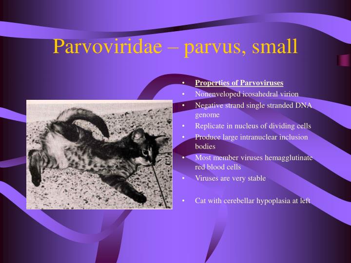
Parvoviridae – parvus, small. Properties of Parvoviruses Nonenveloped icosahedral virion Negative strand single stranded DNA genome Replicate in nucleus of dividing cells Produce large intranuclear inclusion bodies Most member viruses hemagglutinate red blood cells Viruses are very stable

E N D
Parvoviridae – parvus, small • Properties of Parvoviruses • Nonenveloped icosahedral virion • Negative strand single stranded DNA genome • Replicate in nucleus of dividing cells • Produce large intranuclear inclusion bodies • Most member viruses hemagglutinate red blood cells • Viruses are very stable • Cat with cerebellar hypoplasia at left
Parvoviridae • Subfamilies and Genera • Subfamily Parvovirinae • Contains parvoviruses of vertebrates • Genus Parvovirus – • Replicate autonomously • Cannot grow in stationary cells • Replicate in cells undergoing mitosis – do not code for enzymes required for replication, BUT • Rely on the enzymes of the replicating cell to perform this function • Resistance – very stable to environmental conditions – extremes of pH and heat – disinfection DIFFICULT
Diseases caused by Parvovirus • Genus Parvovirus • Virus – Feline parvovirus – Feline panleukopenia • Canine Parvovirus 1 – mild diarrhea • Canine parvovirus 2 - enteritis, leukopenia, myocarditis etc. • Subtypes 2a and 2b • Porcine parvovirus – Reproductive disorders – SMEDI • Mink parvovirus – panleukopenia, enteritis • Pathogenesis - • Determined by the need for mitotic cells for viral replication • Fetus or newborn – Pantropic – Virus may infect a wide range of cells in various tissues and organs • Older neonates – narrower range of cells affected • All age groups – the continuous division of cells of the lymphoid tissue, bone marrow and intestinal epithelium are target sites – leukopenia and enteritis
Feline Panleukopenia • Highly contagious often fatal disease of cats • Most severe in kittens • Synonyms – Feline distemper, feline infectious enteritis • Etiologic agent – feline parvovirus – One serotype • Antigenically related to canine parvovirus 2 and mink parvovirus • Distribution – worldwide • Hosts – all members of the cat family, both domestic and wild are susceptible • Raccoon, mink, coatimundi etc. • Epidemiology – virus is ubiquitous due to its contagious nature and resistance and persistence in the environment
Feline Panleukopenia • Epidemiology continued – • Most subclinical – 75-85% of unvaccinated cats has developed significant antibody titers by one year of age • Unvaccinated kittens are protected through maternal antibodies for up to 3 months • Recovered cats are solidly immune, but can EXCRETE virus in FECES AND URINE FOR 6 WEEKS (OR LONGER) • Extreme resistance of the virus to inactivation and the high rates of viral excretion – 109 the infective dose/ gram of feces – • Results in high levels of environmental contamination – Virus may survive for more than one year in a suitable environment and can be transported via fomites • Owners should NOT adopt an unvaccinated kitten and bring it into the house if parvovirus was ever present
Feline Panleukopenia • Transmission – cats infected ORONASAL route by exposure to infected animals or their secretions – urine, saliva, feces and vomitus or fomites • Mechanical vectors – fleas, humans, flies, etc. • Pathogenesis of Feline Panleukopenia • If exposed in utero – Early fetus will die and be resorbed • Mid-late gestation – abort mummified fetuses • Late gestation – eye, cerebrum and hydranencephaly • **Feline parvovirus produces variable effects on kitttens from the same litter** • Early neonatal – lymphoid tissue affected, cerebellar hypoplasia, bone marrow • Older postnatal – SPF – lymphoid tissue 18-24 hours for replication – adequate titer • Inadequate titer – viremia, 2-7 days in all body tissues either result in DIC and death or recovery
Panleukopenia • Results from destruction of various white blood cell elements – lympocytes, neutrophils, monocytes – including both those present in the circulation and those in the lymphoid organs – • Thymus, bone marrow, spleen, lymph nodes, and Peyer’s Patches • Platelets are also destroyed • Enteritis – • Feline parvovirus multiplies in the rapidly dividing intestinal epithelial cells – crypts of Lieberkuhn. • Normally the epithelial cells at the tips of the intestinal villi are continuously lost into the lumen, replaced by the dividing cells of the crypts • With disease, the villous cells are not replaced, resulting in short non-absorptive cells • Diarrhea and poor absorption results
Central Nervous System Infection and DIC • The CNS, optic nerve, and retina are susceptible to damage by FPV during prenatal or early neonatal development • Cerebellar hypoplasia is the most common • Cerebellar hypoplasia – observed in fetuses infected during the last two weeks of pregnancy and the first two weeks of life • Neonatal cerebellum damaged by FPV is reduced in size and has disordered cell layers • ********************************************************************* • Disseminated intravascular coagulation • Kittens with feline panleukopenia are susceptible to secondary bacterial infection with enteric microflora • Gram negative endotoxemia with or without bacteremia is a common sequelae of systemic FPV infection • DIC results from endotoxic activation of the alternative complement pathway
Feline Panleukopenia and DIC • Clinical signs – most common in kittens 3-5 months of age • Incubation period – 5 days (2-10 day range) • Fever, depression, anorexia, rough coat, repeated vomiting, profuse persistent bloody diarrhea • Dehydration – secondary bacterial infections, and DIC are the major causes of death • Cerebellar hypoplasia. Kitten is permanently ataxic • Usually not apparent until the kitten begins to walk at 3-4 weeks of age • Diagnosis – Clinical signs, hematological examination, and postmortem findings • Virus isolation in cell culture. Swabs from pharynx or rectum, spleen, ileum or mesenteric lymph node • Direct ELISA or immunofluorescence for antigen in tissues • PCR assay for the detection of viral DNA in tissues • Direct hemagglutination of pig or rhesus RBCs by virus present in feces • Paired serum samples. • Virus neutralization test, ELISA, or hemagglutination inhibition test to check for 4 fold increase in antibody titer
Feline Panleukopenia and DIC • Treatment – supportive measures • Good nursing care, fluid therapy, and withholding food in early stages of the disease to lessen vomiting and slow intestinal mitotic activity • Administration of broad-spectrum antibiotics • Control – • Large catteries – strict hygiene and quarantine of incoming animals • Disinfection – virus survives disinfection with 70% alcohol, various dilutions of organic iodines, phenolics, and quaternary ammonium compounds – QUATS • FPV – INACTIVATED BY BLEACH – 6% sodium hypochlorite, 4% formaldehyde, and 1% glutaraldehyde in 10 minutes at ROOM TEMPERATURE • Immunity –neutralizing antibodies can be detected within 3-5 days of infection • Immunity after natural infection appears to be lifelong • Attenuated – modified live-virus vaccine and inactivated vaccine are used • MLV should not be administered to cats that are pregnant, immunosuppressed or sick or to kittens less than 4 weeks old
Canine Parvovirus • Canine Parvovirus disease first recognized in 1978 when it caused a worldwide pandemic – severe gastointestinal illness • Etiologic agent – Canine parvovirus 2 – 2a, 2b • In North America, disease is caused mostly by CPV-2b. Both 2a and 2b are circulating in Europe in other parts of the world • Canine parvovirus 1 – was isolated in feces of dogs in 1967 but is not a major cause of disease • Hosts – domestic and wild canidae – • self-limiting in cats with no clinical signs • Epidemiology – similar to that of feline parvovirus – CPV-2 can persist on fomites for 5 months or longer. • Disease now has lower morbidity and mortality rates as immunity has built up in the dog population
Canine Parvovirus • Transmission –similar to feline panleukopenia • Pathogenesis same as feline, but no cerebellar hypoplasia in puppies • Clinical features – three distinct age-related syndromes have been recognized • 2-12 days – generalized neonatal disease – uncommon • 3-8 weeks – myocarditis –can also develop from infection in utero. Usually all puppies are affected in litter. • Sudden death or a pup may succumb after a short episode of illness • Less common now
Canine Parvovirus • 2-4 months – panleukopenia/ enteritis – most common • Diagnosis – same as feline panleukopenia • Direct ELISA kit for feces detection • IgM capture ELISA used to determine recent infection • Control – same as feline panleukopenia • Immunity – same as feline panleukopenia. Immunity after natural infection appears to be lifelong • Attenuated live and inactivated vaccines available
Porcine Parvovirus - PPV • Porcine Parvovirus infection – 47th day of gestation • Worldwide and endemic in many swine herds • Virus as been isolated in association with respiratory disease and vesicular disease of the feet and mouth in young pigs and neonatal systemic disease • Transmission – oronasal in the dam followed by transplacental transmission • Venereal transmission – possible because infected boars shed virus in the semen for protracted periods
Porcine Parvovirus • Pathogenesis – • virus replicates in lymphoid tissues or organs, salivary glands and other organs • Replicates well in peripheral blood lymphocytes – T and B and NK cells • Reproductive failure is due to exposure of nonimmune females to PPV any time during the first 8 weeks of gestation • Embryo or fetus – less than 30 days – embryo or fetus dies and is resorbed • Early fetus – 30-70 days – fetuses die and become relatively dehydrated – mummified • Late fetus – more than 70 days – develop lesion by nay develop in utero and produce antibody to PPV – immunocompetence of pig fetuses starts at 55-70 days • Boars, sows and gilts – mostly inapparent or subclinical infections • Diagnosis – • Fluorescent antibody staining of frozen sections of fetal tissues results in rapid diagnosis
Porcine Parvovirus • Control – • inactivated and attenuated vaccines are widely used • Nonimmune swine may be exposed to virulent virus several weeks before breeding to establish immunity • E.g. commingling gilts and boars with other swine that may be shedding PPV – like older breeding stock • Unlike most parvoviruses, swine parvovirus causes persistent infection with chronic shedding • SMEDI – other causes include: • Porcine enterovirus 2-11 • Porcine herpesvirus 1 – pseudorabies • Porcine arterivirus – porcine reporductive and respiratory syndrome