Download
1 / 25
490 likes | 1.23k Views
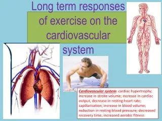
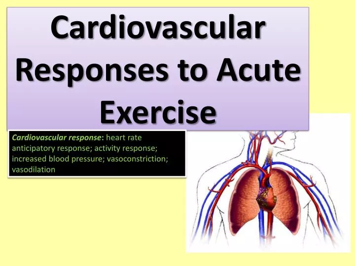
Cardiovascular Responses to Acute Exercise. Cardiovascular response : heart rate anticipatory response; activity response; increased blood pressure; vasoconstriction; vasodilation. The Goal of the CV system is . To meet the increased demands needed to perform exercise

E N D
Cardiovascular Responses to Acute Exercise Cardiovascular response: heart rate anticipatory response; activity response; increased blood pressure; vasoconstriction; vasodilation
The Goal of the CV system is To meet the increased demands needed to perform exercise To meet such demands the following come into play: Heart rate (HR)- beats per minutes Stroke volume (SV) – Amount of blood pumped from the ventricles in one beat Cardiac output (Q) -The amount of blood that is pumped by the heart per unit time, measured in litres per minute (l/min). Blood pressure (BP) - is the pressure exerted by circulating blood upon the walls of blood vessels Blood flow
Resting Heart Rate (RHR) Typically RHR = 60-80 bpm (beats per minute) Pre-exercise HR usually increases above normal resting values This is an anticipatory rise and is not a reliable estimate of RHR - RHR must be taken sometime before exercise Increases in HR are due to the sympathetic nervous system (SNS) releasing adrenaline. Once exercise has started the increase in carbon dioxide (CO) and lactic acid in the body is detected by the chemoreceptors; signals are then sent back to the SNS and more adrenaline is released this further increases HR heart rate increases linearly from about 60 bpm to a maximum of about 200 bpm
Steady-State HR HR increases until it reaches a plateau, when rate of work is held constant at sub-maximal intensity Optimal HR for meeting the circulatory demands at that rate of work The lower the steady-state HR at each exercise intensity, the greater the cardiorespiratory fitness
Maximum HR (MHR) Highest HR value achieved in an all-out effort to the point of exhaustion Remains constant from day to day but decreases with age Approximated by: HRmax= 220-age or HRmax= 208- (0.7 x age)
Stroke Volume (SV) SV is the amount of blood pumped from the ventricles in one beat (specifically the left one) SV is a major determinant of cardiorespiratory endurance capacity at near-maximal and maximal exercise intensities The fitter you are the greater your SV generally! Resting SV is normally around 70-90ml (0.07 -0.09L) In general males have a bigger SV than females SV values are determined by: Volume of venous blood returned to the heart Ventricular dispensability Ventricular contractility Aortic or pulmonary artery pressure
Stroke Volume (SV) stroke volume rises during the initial stages of work and then levels off until near maximal levels where it slightly declines due to decreased ventricular filling time in early exercise the increase is related to increases in both HR and SV. Later increases are due to HR only assuming that SV levels out
Stroke Volume Trained Vs Untrained • Trained individuals have a larger SV then untrained as you can see from the graph!
Increased venous return (preload) (the muscle pump and respiratory pump help with venous return during exercise): extent to which ventricle fills with blood and stretches and subsequently contracts more forcefully: Frank-Starling mechanism Blood is returned through the veins to the heart and enters the atria 2. Blood then moves from the atria to the ventricles causing the myocardium (cardiac muscle) in the ventricles to stretch You can mimic preload by stretching an elastic band, the further the stretch the elastic band the greater the distance it will travel when you release it!!! 3. The greater the venous return the more blood that enters the ventricles, the greater the myocardium is stretched. The further it is stretched the stronger and more forceful the contract will be
The Muscle thoracic Pump Helps Venous Return • During exercise the muscle pump functions to return blood to the heart, or increase venous return; the muscles contract and squeeze the veins to push blood back up to the heart • the thoracic or respiratory pump serves the same function, i.e, as you breath in and out this compresses veins in the chest and abdomen to increase venous return to the heart
What causes SV to increase during exercise? • SV values can also increase due to increased ventricular contractility from neural stimulation from the sympathetic nerve (from the Central Nervous System CNS) • SV values can increase also due to decreased total peripheral resistance in the blood vessels due to vasodilation of blood vessels in exercising skeletal muscle
Cardiac Output (Q) Q is the amount of blood pumped from the heart every minute (litres per minute) and is the product of: HR x SV As HR and SV increase therefore so does Q during exercise, to a maximum! Resting Q is about 5.0 L/min, but does vary with size of person There is a linear relationship between Q and exercise intensity up to 20-40 L/min When level of exercise exceeds 40% to 60% of maximal exercise capacity, SV either plateaus or increases at a much slower rate Further increases in Q at this point are due to increases in HR
Changes in Q (Cardiac Output) and SV As you can see in a trained and untrained individual their SV starts to plateau at a HR of around 120bpm, but Q still increases this is due to increases in HR. However an Elite individuals SV capacity is greater than untrained individuals!
Cardiac Output and Intensity • Here you can see the linear relationship between exercise intensity and Q • This individuals Qmax is around 24L/Min
Changes in HR, SV, and Q with Changes in Posture and Exercise SV changes are due to changes in venous return, ventricular contractility and peripheral resistance HR and activity/exercise intensity have a linear relationship Q and activity/exercise intensity have a linear relationship
Blood Pressure • Cardiovascular Endurance Exercise: • Systolic Blood Pressure (SBP) increases in direct proportion to increase in exercise intensity • As exercise begins the baroreceptors found in the aortic and carotid arteries detect a decrease in blood pressure specifically SBP • The central nervous system (CNS) responds by constricting (vasoconstriction – narrowing of the blood vessel lumen) blood vessels and increasing SBP and further increases HR • Eventually the CNS detects that SBP needs to be reduced and is reduced via the vasodilation of the vessels. The CNS will continue to attempt to regulate BP throughout exercise until maximal levels are reached • Diastolic Blood Pressure (DBP) does not change significantly (may even decrease) • Therefore little change in Mean Arterial Pressure (MAP) which is a product of both SBP and DBP • Resistance Exercise: • Can exaggerate BP as high as 480/350 bpm • Some BP increases can be attributed to the Valsalva maneuver (performed by attempting to forcibly exhale while keeping the mouth and nose closed)
Blood Pressure Response to Exercise Continued McArdle et al., Exercise Physiology, Lippincott, 2001
Blood Pressure Responses • As exercise intensity increases SBP in both arms and legs increases in a linear fashion • Small or little changes in DAP
Blood Flow Acute changes in Q and BP during exercise allow for increased total blood flow to the body. Blood flow patterns change in transition from rest to exercise – blood must be redistributed to other areas such as muscle this is often called a vascular shunt Through Sympathetic Nervous System (SNS), blood is redirected to active areas during exercise SNS activity cause the vasoconstriction (narrowing of vessels) and vasodilatation (widening of vessels) of blood vessels. Also pre capillary sphincters open and close to allow for blood to either travel in or away from a certain area of the body. This causes blood to be redirected to other areas of the body during exercise.
Blood Flow Cont SYSTEMIC BLOOD DISTRIBUTION VASCULAR SHUNT blood is redistributed towards active skeletal muscle during exercise and away from inactive organs as body heat builds up some blood flow is shifted to the skin to help maintain internal temperatures within acceptable limits • VASOMOTOR CONTROL • VASODILATION • dilation of arterioles and opening of precapillary sphincters increases blood flow to active muscle • VASOCONSTRICTION • constriction of arterioles and closure of precapillary sphincters reduces blood flow to inactive organs
Vascular Shunt Below is a diagram showing how pre capillary sphincters along with vasoconstriction and vasodilation help shunt the blood to active areas of the body.
Absolute Relative to total blood volume