Download
1 / 26
270 likes | 554 Views
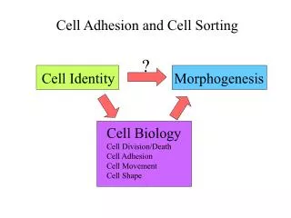
5. Cell adhesion and the actin cytoskeleton. The cell cortex The cytoskeleton networks The actin cytoskeleton Adhesion proteins The extracellular matrix The cell architecture is regulated by the adhesion zones. 1. The cell cortex. Actin microfilaments. 0.2 µm. 5 nm.

E N D
5. Cell adhesion and the actin cytoskeleton The cell cortex The cytoskeleton networks The actin cytoskeleton Adhesion proteins The extracellular matrix The cell architecture is regulated by the adhesion zones 1
The cell cortex Actin microfilaments 0.2 µm 5 nm Medalia et al. 2002 Science 298: 1209-1213 100 nm cryoelectron tomography Protein complexes membranes 2
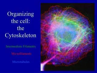
The cell cytoskeleton networks Cells possess two or three cytoskeleton networks made of polymerized proteins : an actin cytoskeleton (microfilaments), a tubuline one (microtubules) and intermediate filaments • The cycle of microfilament polymerisation-depolymerisation is coupled to ATP hydrolysis by actin • The cycle of microtubule polymerisation-depolymerisation is coupled to GTP hydrolysis by tubulin • Intermediate filaments are controlled by protein phosphorylation and dephosphorylation About hundred proteins control the growth and dissociation rates or are associated to microfilaments and microtubules : actin binding proteins, microtubule associated proteins, bundling proteins, molecular motors ... Intermediate filaments 3 microfilaments microtubules
The actin cytoskeleton Microfilaments are actin polymers, an ATP binding and ATP hydrolysing protein. The cycle of microfilament polymerisation-depolymerisation is coupled to ATP hydrolysis by actin Microfilaments constitute the membrane cortex which allows the deformation of the plasma membrane. Stress fibers are actin microfilaments that link adhesion focal points. Actin microfilaments are reversibly linked to the plasma membrane by specific proteins (talin, catenin, ezrin ...) Mechanical forces are exerted by actin polymerization at the plasma membrane and between actin microfilaments by molecular motors reflection interference contrast microscopy actin immunofluorescence 4
Structure of actin microfilaments barbed end pointed end 4 nm 5
Actin polymerization kinetics : 1) no hydrolysis At steady state, V = (1/a)dL/dt = konC – koff Critical concentration : Cc = koff/kon = Keq Cc+end = Cc-end V (polymerization rate) + end - end C (monomer concentration) Cc 6
ATP hydrolysis Actin polymerization kinetics : 2) ATP hydrolysis V (polymerization rate) + end + end - end - end AATP AADP C (monomer concentration) CcATP CcADP 2 sec Pi At steady state, during treadmilling V = VT(+) + VD(-) = konT.CT – koffT + konD.CD – koffD CD~ 0.1 CT , koffT < koffD and konD < konT In steady state CTc koffD/konT 7
Experimental polymerization dynamics Fujiwara et al. PNAS 2007 104 : 8827–8832 • CTc koffD/konT 0.25/11.6 = 0.02 µM • Physiological actin concentration : about 100 µM • Most actin is complexed to thymosin [ActinT] 10 µM • The theoretical actin microfilament elongation rate is : • dL/dt = a.VT = a.konT.[ActinT] < (4 nm).(12 µM-1.s-1).(10 µM) = 0.48 µm.s-1 • At the pointed end : • dL/dt = a.VD = a.koffD = (4 nm).(0.25 s-1) = 1 nm.s-1 • To be compared to the actual velocity of cell edge movements : up to 10 µm.s-1 8
In vivo evidence of microfilament treadmilling by FRAP experiments Injection of rhodamine-actin Localized photobleaching Transport (treadmilling ) 9
How cytoskeleton molecules exert forces ? 1. Actin polymerization : the elastic brownian ratchet model (Mogilner and Oster 1996 Biophys. J. 71 : 3030-45) Stall force Mechanical work = ATP hydrolysis F.d = DG/N = DG° + RT ln{[actinATP]} = kBT ln{kon[actinATP]/koff} actinATP + filamentN filamentN+1 + Pi 10
How cytoskeleton molecules exert forces ? 2. Actin microfilament pressure at nucleation zones (C Sykes & J Prost (2005) P.N.A.S., 102, 7847-52) • Actin nucleation at the bead surface • Concentric growth of actin microfilaments • Shear stress accumulation • Breaking the actin gel, resulting in asymmetric growth 11
Visualization of F-actin network movement in motile keratocytes with FSM Yam et al. JCB 178:1207-1221, 2007 30x real time 12 12 Actin filament dynamics
How cytoskeleton molecules exert forces ? 3. Molecular motors sliding on microfilaments 13
Regulation of cytoskeleton dynamics ↑ Stress fibers ↑ Lamellipodia ↑ Filopodia Ridley & Hall 1992 Allen et al. 1997 14
Example : microvilli at the plasma membrane Brush border cells of the intestinal epithelium Actin cytoskeleton Plasma membrane 15
Cell adhesion and the actin cytoskeleton The cell cortex The cytoskeleton networks The actin cytoskeleton Adhesion proteins The extracellular matrix The cell architecture is regulated by the adhesion zones 16
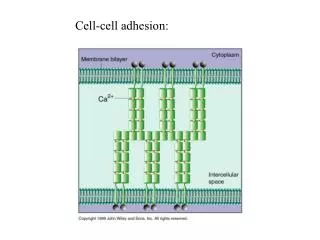
The extracellular matrix and cell adhesion molecules The extracellular matrix is made of macromolecules (proteins and polysaccharides) synthesized and secreted by cells. It provides a specific mechanical and chemical environment for the cells. Cell adhesion molecules are proteins expressed at the surface of cells that mediate binding to other cells or to the extracellular matrix. 17
Extracellular matrix proteins have a complex structure • Proteins of the ECM have a very large multi-domain structure • They often contain growth factor-like domains or bind growth factors A) Fibronectin. Encoded by a single gene but alternatively spliced at three regions [blue circles and box and V (variable) segment] to generate 12 proteins in rodents and 20 in humans. FN3 domains are widespread in ECM proteins. Binding sites for other matrix proteins are marked. The heparan sulfate–binding site can interact with proteoglycans or with syndecan, an integral-membrane proteoglycan. Integrin-binding sites; RGD (indicated by an asterisk) and LDV (Leu-Asp-Val, indicated by a pound sign). FN is a proangiogenic molecule, whose function depends on both the RGD site and the two alternatively spliced FN3 domains. FN also binds the proangiogenic growth factors VEGF and HGF. B) Fibrillin-1. Fibrillins include EGF-like domains, found in many ECM proteins, as well as TB (TGFb-binding, denoted by T) and hybrid (H) domains, specific to fibrillins and LTBPs. Binding sites for other matrix proteins and growth factors are marked. C) LTBP-1. Four-gene family with structures related to fibrillins. Known binding sites for TGF-b/LAP latent complex (SLC, blue), fibrillin, and FN are marked. RGD (asterisk) sequences in fibrillins and LTBPs may bind integrins. D) Thrombospondin-1 (TSP-1). TSPs contain TSP1 repeats (also found in other ECM proteins), EGF-like repeats, and a VWC domain, known in other proteins to bind BMPs. TSP3 repeats (purple) and C-terminal domains are unique to TSPs and bind multiple Ca2+ions. The RGD (asterisk) sequence is known to bind to integrins. TSPs 1 and 2 have the structure shown, and both have antiangiogenic activity located in the TSP1 repeats, which bind to the CD36 receptor (39) RO Hynnes (2009) the extracellular matrix : not just pretty fibrils Science 326 : 1216-19
Madin-Darby Canine Kidney cells, an epithelium model Polarized cell : two compartments around the cell 22
Tight junctions separates apical from basolateral compartments Electron micrographs of cells in an epithelium in which a small, extracellular, electron-dense tracer molecule has been added to either the apical side (on the left) or the basolateral side (on the right). In both cases, the tracer is stopped by the tight junction. (photo Daniel Friend) The sealing strands hold adjacent membranes together. They are composed of transmembrane proteins (claudins, occludins) that make contact across the intercellular space and create a seal. 23
The cell architecture is regulated by the adhesion zones http://www.cytoo.com/ 24
Golgi apparatus (in red) in RPE1 cells in standard culture conditions Golgi apparatus (in red) in RPE1 cells on fibronectin micropatterns (in green) 26