Download
1 / 63
630 likes | 661 Views
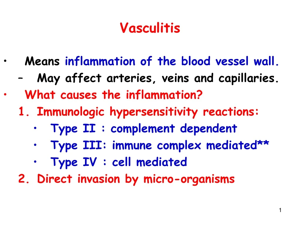
Vasculitis. Means inflammation of the blood vessel wall. May affect arteries, veins and capillaries. What causes the inflammation? Immunologic hypersensitivity reactions: Type II : complement dependent Type III: immune complex mediated** Type IV : cell mediated

E N D
Vasculitis • Means inflammation of the blood vessel wall. • May affect arteries, veins and capillaries. • What causes the inflammation? • Immunologic hypersensitivity reactions: • Type II : complement dependent • Type III: immune complex mediated** • Type IV : cell mediated • Direct invasion by micro-organisms
Etiopathogenesis Immunologic mechanisms • Immune complexe deposition • Responsible for most cases*** • Deposition of immune complex • Activation of complement • Release of C5a • C5a chemotactic for neutrophil • Neutrophils damage endothelium and vessel wall fibrinoid necrosis. • Endothelial damage thrombosis • Ischemic damage to tissue involved. • Example of IC mediated Vasculitis = Henoch-Schonlein purpura
Etiopathogenesis Immunologic mechanisms • Type IV hypersensitivity: delayed type of hypersensitivity reaction • implicated in some types of vasculitis due to presence of granulomas. • Example: Temporal arteritis • Direct Invasion: • by all classes of microbial pathogens • Rickettsiae • Meningococcus • Fungus
Laboratory testing in vasculitis • Antineutrophil cytoplasmic antibodies (ANCA) • Erythrocyte sedimentation rate (ESR)
Antineutrophil cytoplasmic antibodies (ANCAs) • Are seen in some types of vasculitis esp small vessel vasculitis • Are circulating ab reactive with neutrophil cytoplasmic ag = ANCA. • The ANCAs activate neutrophils • Cause release of enzymes and free radicals resulting in vessel damage. • ANCA titers correlate with disease activity. • Detected by immunofluorescence
Two types of ANCAs • Cytoplasmic (c-ANCAs): • Ab directed against proteinase 3 in cytoplasmic granules. • Cytoplasmic staining pattern • Example:Wegener’s granulomatosis. • Perinuclear (p-ANCAs): • Ab directed against myeloperoxidase. • Perinuclear pattern of staining • Example:Churg-Strauss syndrome, PAN.
Classification of Vasculitis : based on vessel size • Large vessel Vasculitis: • Giant cell arteritis * • Takayasu’s arteritis * • Medium vessel Vasculitis • Polyarteritis nodosa (PAN)* • Kawasaki’s disease* • Thromboangitis obliterans (TAO)* • Small vessel Vasculitis • Hypersensitivity vasculitis • Henoch Schonlein purpura* • Churg Strauss syndrome • Wegener granulomatosis *
Clinical manifestations of vasculitis • Clinical picture depends on the size and extent of the vessel involvement. • Large vessel Vasculitis: • Presents with loss of pulse or • Stroke • Medium vessel Vasculitis • Presents with infarction or aneurysm • Small vessel Vasculitis • Presents with Palpable purpura* • General features: • Fever, weight loss, malaise, myalgias
Patient Profile # 1 • Old female patient presents with • Headache in the temporal region • Pain in the jaw while chewing • Muscle aches and pains • Develops problems with vision. • On examination: • Has nodular and palpable temporal artery. • Labs: • elevated ESR • Biopsy: ( temporal artery) • granulomatous inflammation with giant cells • Diagnosis: • Giant cell (temporal) arteritis
Large vessel vasculitis Giant cell (temporal) arteritis • Is the most common vasculitis**. • Occurs in women > 50 years (Female > male) • Vessel involvement:: • Typically involvestemporal artery and extra-cranial branches of external carotid. • Involvement of ophthalmic branch of external carotid blindness. • Etiopathogenesis: • Type IV hypersensitivity mediated reaction causing granulomatous inflammation.
Giant cell arteritis: Pathology • Affected vessel are cordlike and show nodular thickening. • Microscopy: • Focal Granulomatous inflammation of temporal artery • Fragmented internal elastic lamina • Giant cells.
Temporal (giant cell) arteritis Giant cell
Giant cell (temporal) arteritis • Clinical features: • Fever, fatigue, weight loss • Temporal headache* (MC symptom), facial pain. • Painful, palpably enlarged and tender temporal artery* • Generalized muscular aching and stiffness (shoulders and hip) • Temporary / permanent blindness*
Investigations: ESR: screening test of choice ; markedly elevated. Temporal artery biopsy : definitive diagnosis (positive in only 60% cases) Treatment: Corticosteroids (to prevent blindness) Giant cell (temporal) arteritis
Patient profile # 2 • Middle aged Asian womanpresents with: • Visual disturbances • Marked decrease in blood pressurein upper extremity and • Absent radial, ulnar and carotid pulses. • Angiography shows: • Marked narrowing of aortic arch vessels • Biopsy: • Granulamatous inflammation with giant cells • Diagnosis: • Takayasu’s arteritis (pulseless disease)
Takayasu’s arteritis (pulseless disease) • Is an inflammatory disease of vessels affecting • the aorta and its major branches • Seen in Asian women <50 years old. • Vessel involvement: • Typically involves the aorta* and the aortic arch vessles* (carotids, subclavian). • Can also involve: pulmonary, renal, coronary • Etiopathogenesis: • Type IV hypersensitivity reaction causing granulomatous inflammation (granulomatous vasculitis)
Takayasu’s arteritis (pulseless disease) • Pathology: • Thickening of vessels ( aorta & branches) with narrow ( stenosis) lumen • decreased blood flow • Microscopic • Similar to/indistinguishable from Giant Cell Arteritis
Takayasu’s arteritis (pulseless disease) • Clinical: • Dizziness,syncope. • Absent upper extremity pulse (pulseless disease)** • Blood pressure discrepancy* between extremitis : low in upper and higher in lower • Visual disturbances • Diagnosis: • angiography
Patient profile # 3 • Young maleIV drug abuser with history of Hepatitis (HBV) presents with • Hypertension, abdominal pain, melena,muscle aches and pains and skin nodulations. • Biopsy of skin nodules: • Segmental transmural inflammation of blood vessels with fibrinoid necrosis. • Labs: • HBsAg +ve • pANCA +ve • Diagnosis: • Polyarteritis nodosa (PAN)
Polyarteritis nodosa (PAN) • A systemic disease. • Vessel involvement: • Affects medium sized & small muscular arteries*. • Typically involves vessels of • Kidney, heart, liver, GIT and skin • Spares the lung** • Etiology: • Mediated by type III hypersensitivity ( ag-ab complex deposition). • Associations: • strong association with HBV antigenemia • hypersensitivity to drugs (IV amphetamines). • Pathogenesis: • immunecomplex deposition (e.g. HBsAg / anti- HBsAg)
Small to medium sized muscular arteries IMMUNECOMPLEX DEPOSITION Activation of complement system Acute inflammation • Damage to vessel wall • neutrophil infiltration • fibrinoid necrosis Thrombosis Aneurysms Infarction in involved organs Nodules
Neutrophils fibrinoid necrosis
PAN • Pathology: • Transmural inflammation (involving all layers). • Lesion in the vessel wall may • involve entire circumference or part of it • Fibrinoid necrosis • Consequences: • development of • Thrombosis infarction • Weakening of vessel wall Aneurysms (kidney, heart and GI tract)
PAN: Clinical features • More common in young to middle aged men • Signs and symptoms: due to ischemic damage. • Target organs: • Kidneys : Vasculitis/infarction hypertension , hematuria, albuminuria. • GI tract: Bowel infarction abdominal pain, melena. • Skin: Ischemic ulcers and nodules. • Coronary arteries: aneurysms, MI • Systemic manifestation: fever, malaise and weight loss. • Cause of death: Renal failure MC COD
PAN • Laboratory findings: • HbsAg positive in 30% of cases • Hematuria with RBC cast • Diagnosis: • arteriography or biopsy of palpable nodulations in the skin or organ involved . • Treatment: • Untreated cases: almost fatal • Good response to immunosuppressive therapy.
Churg-Strauss Syndrome (Allergic granulomatous angitis) • Is a systemic vasculitis that occurs in persons with asthma*. • A variant of PAN. • Involves small* & medium vessels of • upper/lower respiratory tract* • heart, spleen, peripheral nerves, skin , kidney. • Pathology: • Inflammation of vessel wall (eosinophils) • Fibrinoid necrosis • Thrombosis and infarction
Churg-Strauss Syndrome (Allergic granulomatous angitis) • Features very similar to PAN but patients with CSS have: • History of atopy • Bronchial asthma, allergic rhinitis and • peripheral blood eosinophilia. • Microscopy: • Similar to PAN • Labs: • peripheral eosinophilia , high serum IgE, • p-ANCA*
Patient profile # 4 • A 4 year old Japanese child presents with • Fever, redness of eyes and oral cavity • Swollen hands and feet • Rash over the trunk and extremities • Peeling of skin and • Cervical lymphadenopathy. • Labs: • ECG changes consistent with myocardial ischemia • Diagnosis: • Kawasaki Disease (mucocutaneous lymphnode syndrome)
Kawasaki’s disease • Is also known as mucocutaneous lymphnode syndrome. • Is an acute self limited febrile illness of infants and children (< 5 yrs). • Is endemic in Japan , Hawaii • One of the manifestations is vasculitis (coronary artery). • In other words: • KD is a childhood vasculitis that mainly targets coronary arteries. • Coronary artery involvement: • can lead to coronary thrombosis or aneurysm formation and its rupture.
Coronary artery aneurysms
Clinical features : Kawasaki’s disease Oral Erythema Conjunctivitis PalmerErythema
Clinical features : Kawasaki’s disease Rash Desquamation Edema: feet and arms
Clinical findings: • High fever • Erythematous rash of trunk and extremities with desquamation of skin. • Mucosal inflammation : cracked lips, oral erythema • Erythema, swelling of hands and feet. • Localized lymphadenopathy (cervical adenopathy) • MCC of an acute MI in children****** • Lab: • Neutrophilic leukocytosis • Thrombocytosis : characteristic finding • High ESR • abnormal ECG (e.g. acute MI)*****
Patient profile # 5 • A young smoker male patient from Israel presents with C/O • Pain in the foot • Which is severe and present even at rest • On examination: • Presence of ulcers and blackish areas over the fingers and toes. • Some missing digits. • Biopsy from lower limb vessel: • Acute inflammation of vessel wall with Obliteration of vessel lumen by a thrombus. • Diagnosis:Thromboangitis Obliterans (Buerger’s Disease)
Buerger’s Disease • Also known as Thromboangitis Obliterans. • Is a peripheral vascular disease of smokers. • Pathology: • Earliest change: Acute inflammation involving the small to medium sized arteries in the extremities (tibial, popliteal & radial arteries). • Inflammation of vessel thrombus formation obliterates lumen ischemia gangrene of extremity. • Inflammation also extends to adjacent veins and nerves. • Involvement of entire neurovascular compartment.
Buerger’s Disease • Clinical findings: • Young-middle age, male, heavy smoker* • Israel*, Japan, India. • Symptoms start between 25 to 40 years • Early manifestation: • Intermittent Claudication in feet or hands • Cramping pain in muscles after exercise, relieved by rest • Late manifestation: • Painful ulcerations of digits • Gangrene of the digits often requiring amputation.
Buerger’s Disease • Diagnosis: • biopsy • Rx: • early stages of vasculitis frequently cease on discontinuation of smoking.
Small vessel vasculitisHypersensitivity (leukocytoclastic) vasculitis • Refers to a group of immune complex mediated vasculitides. • Characterized by: • Acute inflammation of small blood vessels • Manifesting as palpable purpura***. • Organs involved: • Usually skin ( other organs less commonly affected).
Hypersensitivity (leukocytoclastic) vasculitis • May be precipitated by • Exogenous antigens • Drugs • E.g. aspirin/penicillin/thiazide diuretics • Infectious organisms • E.g. strep/staph infections,TB,viral diseases • Foods • Chronic diseases • E.g. SLE, RA etc.
Hypersensitivity (leukocytoclastic) vasculitis • Pathology: • acute inflammation of small blood vessels (arterioles, capillaries, venules) • Neutrophilic infiltrate in vessel wall. • Leukocytoclastic refers to nuclear debris from disintegrating neutrophils • The neutrophils undergo karyorrhexis. • Erythrocyte extravasation