Download
1 / 9
90 likes | 276 Views
Tertiary protein structure viewing and prediction. July 3, 2007 Learning objectives- Learn how to manipulate protein structures with Deep View software. Learn the steps to protein structure modeling with Deep View. Workshop-Manipulation of the lysozyme and hemoglobin.

E N D
Tertiary protein structure viewing and prediction • July 3, 2007 • Learning objectives- Learn how to manipulate protein structures with Deep View software. Learn the steps to protein structure modeling with Deep View. • Workshop-Manipulation of the lysozyme and hemoglobin.
Protein structure viewers • RasMol • Deep View • Cn3D • WebLabViewer • Chimera
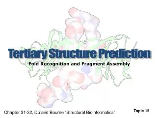
Steps to tertiary structure prediction • Comparative protein modeling • Extrapolates a new structure based on solved structures that are related by sequence. • Steps for SWISS-Model • Identification of modeling templates. • Alignment between target and templates. • Model building. • Evaluation.
Step 1: Identification of modeling templates • One chooses a cutoff value from FastA or BLAST search ( E<10-5) and perform BLAST search of Protein Data Bank. • Up to ten structure templates can be used but the one with the highest sequence similarity to the target sequence (lowest E-value) is designated as the reference template. Its structure is given the most weight. • Ca atoms of the templates are selected for superimposition. • This generates a structurally corrected multiple sequence alignment
Step 2: Alignment • Up to 5 template structures are superimposed. • Incompatible templates are removed. • Pairwise alignment is created between target and main template structures.
Step 3: Building the model • Framework construction • Average the position of each atom in target sequence, based on the corresponding atoms in template (start with C atoms) • Each loop is defined by the length of the • loop and C atom coordinates of the four residues preceding • and following the loop. Constraint space programming is used. • Portions of the target sequence that do not match the • template are constructed from a “spare part” algorithm.
Step 3: Building the model • Completing the backbone-a library of PDB entries is consulted to add carbonyl groups and amino groups. The 3-D coordinates come from a separate library of pentapeptide backbone fragments. These backbone fragments are fitted onto the target C alpha carbons. The central tri-peptide atoms are averaged from each backbone atom (N,C,C(O)). • Side chains are added from a table of most probable rotamers given a certain backbone conformation.
Step 4: Energy Minimization Model refinement-minimization of energy (GROMOS96 force field)