Download
1 / 21
210 likes | 291 Views
Research Training. Yu Xiao Di Uppsala University. Chaperone.

E N D
Research Training Yu Xiao Di Uppsala University
Chaperone • A protein that helps other proteins to fold.Molecular chaperones do not specify the tertiary structure of a protein, they merely help the protein find that correct structure. They prevent protein aggregation by holding the protein in an open conformation until it is completely synthesized and ready to fold.
Our task • Our task is to understand the structure of virulence organelles. We decided to determine a minimal fiber of F1 capsular antigen. This fiber is a complex containing one molecule of the Caf1M chaperone and two molecules of Caf1 subunit (Caf1M-Caf1’-Caf1’’).
Protein expression • Host strain E. coli DH5α strain was transformed with pFM1-1-T7F. For expression of mutated complexes, cells were grown to an OD600nm of 0.8-1 in LB medium ( Maniatis et al., 1998) containing 60 mg L–1 ampicillin. Protein expression was induced with 0.3-0.4 mM IPTG for 2 h.
Purification complex • Samples were loaded onto an 8 ml Mono Q column. Fractions were analyzed by isoelectric focusing ( IEF) on pH 4-6 gradient gels to detect the target complex. Samples were concentrated and incubated with TAGZym at 33°overnight. Digestion efficiency was checked by IEF. Samples were dialysis against 40 mM NaAC, pH 4.6-4.7 and loaded into a 1 ml Mono S equilibrated column with 40 mM NaAC, pH 4.6-4.7 . Sample was concentrated to 30 mg/ml on a 2 ml VivaSpin device with MW cut off of 10 kDa, 2050 rpm.
Crystallization • Crystallization was performed by the hanging-drop vapour-diffusion method at 293 K. In all experiments the crystallization drops, containing 1.5 µl protein solution (diluted) and 1.5 µl precipitant solution, were equilibrated against 1 ml precipitant solution. The initial crystallization conditions were found using Hampton Crystal Screen. Small crystals were found in condition #46. Best crystals were found in droplets containing 10 % PEG 8000, 0.5 M NaAC, 0.1 M Ca(AC)2, pH 5.7-6.0.
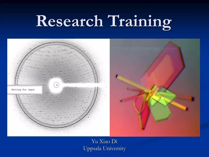
X-ray diffraction study • Diffraction data were collected under liquid-nitrogen cryoconditions at 100 K. To avoid damage on freezing, crystals were soaked for 10-30 s in a cryoprotection solution containing 14 % PEG 400, 11 % PEG 8000, 60 mM NaAC, 120 mM Ca(AC)2, pH 5.7-6.0. Crystals were flash-cooled by rapidly moving them into the cold nitrogen stream or by dipping them in liquid nitrogen. X-ray diffraction data were collected using rotating anode as an X-ray source (λ=1.45 Å) and recorded on a Mar-345 detector. Images of the diffraction of the crystals were analyzed to find preliminary space group to be either P222 or P212121 or P21212 or P2221 with cell unit a=71.2 Å, b=96.5 Å, c=166.4 Å, α=β=γ =90°. It was predicted that asymmetric unit contains two molecules.
Wake up ! Story is not ending • Complete diffraction data set using one of the frozen crystals will be collected at synchrotron in France. And then the structure of the protein complex will be solved by molecular replacement (MOLREP). Later, other characters of this complex and another mutated protein complexes will be studied.
Acknowledgments • I would like to thank Stefan Knight for letting me do my research training in his group. I also want to thank my supervisor Anton Zavialov for helping me solve lots of problems.
Reference • Anton V. Zavialov, Jenny Berglund (2003). Structure and biogenesis of the capsular F1 Antigen from Yersinia pestis: Preserved Folding Energy Drives Fiber Formation. Cell 113:587-596. • A. V. Zavialov, J. Kersley (2002). Donor strand complementation mechanism in the biogenesis of non-pilus systems. Molecular microbiology 45(4): 983-995. • Frederic G. Sauer, Stefan D. Knight (2000). PapD-like chaperones and pilus biogenesis. Cell & Developmental biology 2000:pp.27-34. • Anton V. Zavialov, Vladimir M. Tischenko (2005). Resolving the energy paradox of chaperone/usher-mediated fibre assembly.Biochem.J.389:685-694. • Devapriya Choudhury, Andrew Thompson (1999). X-ray Structure of the FimC-FimH Chaperone-Adhesin Complex from Uropathogenic Escherichia coli. Science 285:1061-1066. • Stefan D Knight, Jenny Berglund (2000). Bacterial adhesins: structureal studies reveal chaperone function and pilus biogenesis. Chemical Biology 4:653-660. • Frederic G Sauer, Michelle Barnhart (2000). Chaperone-assisted pilus assembly and bacterial attachment. Structural biology 10:548-556.
Good Luck in the exam coming next week !!! • Thank you. • 12-17-2005