Download
1 / 21
210 likes | 530 Views
Protein Metabolism. CH353 April 3, 2008. Genetic Code. Codons of genetic code are in 5 ′ → 3 ′ direction on mRNA Initiation codon (AUG) is green 3 termination codons are red (UAA, UAG, UGA) 2 amino acids have unique codons (Trp - UGG, Met - AUG)

E N D
Protein Metabolism CH353 April 3, 2008
Genetic Code • Codons of genetic code are in 5′→3′ direction on mRNA • Initiation codon (AUG) is green • 3 termination codons are red (UAA, UAG, UGA) • 2 amino acids have unique codons (Trp - UGG, Met - AUG) • Most codons are degenerate – more than one codon for the same amino acid • 9 amino acids have 2 codons; 1 has 3 codons; 5 have 4 codons; 3 have 6 codons
Codon – Anticodon Recognition • Wobble allows one tRNA (anticodon) to recognize multiple codons • Base selection at 1st position of anticodon allows base-pairing with multiple bases in 3rd position of codon • Wobble permits recognition of all 61 codons with only 32 tRNAs (including unique initiator tRNA)
A ribozyme for synthesizing proteins 50S and 30S subunits, with mRNA, with/without tRNAs bound at the E, P and A sites The Ribosome
Composition and Mass of Ribosomes • Ribosomal subunits are named by their sedimentation velocities (S) • rRNA forms the core of ribosomal subunits; proteins are secondary surface elements • rRNA catalyzes peptide synthesis; no protein within 18 Å of active site • 16S rRNA, tRNAs, mRNA cooperate for correct placement of amino acids • Eukaryotic Ribosomes are ~56% larger than prokaryotic ribosomes • Eukaryotic ribosomes have more proteins, larger rRNAs and an extra rRNA in 60S subunit
Cloverleaf pattern of Watson-Crick base-pairing in tRNA, highlighting typical or invariant nucleotides 3D stucture of tRNA has ‘L’ shape; stacking anticodon arm + D arm, and TyC arm + amino acid arm tRNA Secondary and Tertiary Structure
Major Stages of Protein Synthesis • Activation of Amino Acids • activate carboxyl group of amino acid for peptide synthesis • establish link between amino acid and biological information • Initiation of Translation • join initiation codon of mRNA to ribosome and initator tRNA • Elongation of Translation • recruit aminoacyl-tRNA based on codon of mRNA template • form peptide bond and advance to next codon • Termination of Translation • recruit termination factors to stop codon • release completed polypeptide • Folding and Post-translational Processing • fold into active form; proteolytic processing; modify amino acids
Aminoacyl-tRNA Synthetases catalyze at least 2 reactions: amino acids activation with ATP, and aminoacyl transfer to tRNA 2 classes of aminoacyl-tRNA synthetases: Class I: Arg, Cys, Gln, Glu, Ile, Leu, Met, Trp, Tyr, Val Class II: Ala, Gly, Asn, Asp, His, Lys, Phe, Pro, Ser, Thr Overall Reaction: ∆G′º = -29 kJ/mol Amino acid + tRNA + ATP → aminoacyl-tRNA + AMP + PPi PPi + H2O → 2 Pi Stage 1: Aminoacylation of tRNA
Class I aminoacyl-tRNA synthetases Initial esterification on 2′ OH Class II aminoacyl-tRNA synthetases Initial esterification on 3′ OH Aminoacylation of tRNA
Proofreading by Aminoacyl-tRNA Synthases • Correct connection between amino acid and anticodon needs to be made here (no amino acid proofreading on ribosome) • Interaction with amino acid at 2 levels of specificity; binding to amino acid and to aminoacyl-AMP • Provides multiplicative lower error rate of ~10-4 • Interaction with tRNAs: multiple tRNAs but one synthetase per amino acid; specificity determined by characteristic nucleotides and structure of tRNAs
Stage 2: Translation Initiation • Step 1: • 30S subunit binds to IF-1 and IF-3 and then to mRNA (AUG at P site) • IF-1 blocks A site, IF-3 prevents premature association with 50S subunit • Step 2: • IF-2–GTP binds to fMet-tRNAfMet, which facilitates binding to 30S complex • tRNA anticodon pairs with codon of mRNA • Step 3: • GTP is hydrolyzed; IF-1, -2, -3 dissociate • 30S complex combines with 50S subunit to form 70S initiation complex
3’ End of 16S rRNA 3’ A U U C C U C C A . . . | | | | | | | | | Translation Initiation on Bacterial mRNA • Correct positioning of 30S ribosomal subunit at initiation codon requires an upstream Shine-Dalgarno sequence, complementary to 3′ end of 16S rRNA • Extent of complementarity and spacing relative to initiation codon determine initiation efficiency; requirements are species specific (more is not always better) • AUG is not always initiation codon (GUG and UUG can be used), but fMet-tRNAfMet is used regardless of codon
Translation Initiation on Eukaryotic mRNA • Initiation codon is nearly always the 5′-terminal AUG • Kozak sequence facilitates initiation (5′) ACCAUGG (3′) • 5′-terminal cap and 3′-terminal poly(A) are essential • eIF4F complex (4A, 4B, 4E, 4G) binds to cap and to eIF3 • Scanning for AUG requires ATP and eIF4A (helicase) • eIF2 similar role as IF-2, binding Met-tRNAiMet (GTP required) • eIF5 involved in assembly of 80S ribosome (GTP required)
Stage 3: Translation Elongation • Step 1: Binding of Aminoacyl-tRNA • aminoacyl-tRNA binds to EF-Tu–GTP • resulting assembly binds to A site of 70S initiation complex • GTP is hydrolyzed; EF-Tu is released; EF-Tu–GTP regenerated with GTP and EF-Ts Proofreading of codon-anticodon interaction: • aa-tRNA complexes with EF-Tu-GTP and with EF-Tu-GDP exist for few milliseconds • If correct codon-anticodon is not found during this time, complex dissociates • 18S rRNA involved in correct base pairing • eEF1a and eEF1bg are eukaryotic analogs
Stage 3: Translation Elongation • Step 2: Peptide Bond Formation • Transfer of the acyl peptide at the P site to the aminoacyl-tRNA at the A site • Nucleophilic attack by amino group (A site) on acyl group (P site) with tRNA as leaving group (remaining bound to P site) • Reaction catalyzed by 23S (or 28S) rRNA • Step 3: Translocation • Ribosome shifts one codon toward the 3′ end of the mRNA (peptidyl-tRNA to P site and empty tRNA to E site) • EF-G–GTP binding to A site mimics the EF-Tu-tRNA complex displacing peptidyl-tRNA • eEF2 is eukaryotic factor analogous to EF-G
Stage 4: Translation Termination • 3 Termination factors (release factors) proteins RF-1, RF-2 and RF-3 facilitate: • hydrolysis of terminal peptidyl-tRNA bond • release of polypeptide and tRNA • dissociation of 70S ribosome into subunits • RF-1 binds UAG or UAA; • RF-2 binds UGA or UAA • RF-1 or RF-2 binds to stop codon, causing peptidyl transferase ribozyme to use water instead of aminoacyl-tRNA as nucleophile (mimic tRNA structure – like EF-G) • RF-3 may function in ribosome dissociation • eRF performs all functions in eukaryotes
Polysomes (Poly-ribosomes) Multiple ribosomes simultaneously translating a single mRNA provides: • Efficient protein synthesis • 10 to 100 ribosomes producing protein from each mRNA • Stability of mRNA • Close spacing of ribosomes protect mRNA from ribonucleases Polysomes occur in both prokaryotic and eukaryotic translation
Coupled Transcription and Translation • Eukaryotic transcription occurs in nucleus but translation occurs in cytosol – requires translocation of processed mRNA to cytosol • Prokaryotes have no nuclei so the nascent mRNA can be translated while it is being transcribed • Eukaryotic mRNA nearly always encode 1 protein (moncistronic) • Prokaryotes typically have multiple coding regions in a single mRNA • Operons transcribe multiple coding regions into one mRNA Operons often coordinate the biosynthesis of proteins serving one function, e.g. multisubunit proteins or metabolic pathways
Stage 5: Post-Translational Processing Cytoplasmic Modifications • Proteolytic processing • hydrolysis of N- and C-terminal amino acids; deformylating fMet • Amino acid modification • phosphorylation, methylation, acetylation • Attachment of lipids • isoprenylation, N-myristoylation, C-palmitoylation • Attachment of prosthetic groups • activation of enzymes and proteins (e.g. coenzymes, heme)
Stage 5: Post-Translational Processing Secretion and Targeting • Signal (or target) peptide removal • hydrolysis of signal or target peptide during secretion • Attachment of carbohydrate groups and lipids • O-linked (Ser or Thr) and N-linked (Asn) glycosylations • C-terminal GPI (glycosyl phosphodylinositol) anchors • Disulfide bond formation • Disulfide-bonded proteins are usually lumenal or extracellular • Proteolytic Processing • activation of pro-enzymes and pro-hormones • Amino acid modification • Glu g-carboxylation, Pro hydroxylation, others
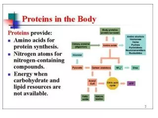
Processes Affecting Protein Levels • Synthesis of primary RNA transcript (transcription) • Posttranscriptional modification of mRNA • Degradation of mRNA • Protein synthesis (translation) • Posttranslational modification of proteins • Protein targeting and transport • Protein degradation