Download
1 / 72
730 likes | 750 Views
Body Fluids & Blood. Movement of water between compartments. Normally, cells neither shrink or swell because intracellular and interstitial fluids have the same osmolarity Increasing osmolarity of interstitial fluid draws water out of cells and cells shrink

E N D
Body Fluids & Blood University of Jordan
Movement of water between compartments • Normally, cells neither shrink or swell because intracellular and interstitial fluids have the same osmolarity • Increasing osmolarity of interstitial fluid draws water out of cells and cells shrink • Decreasing osmolarity of interstitial fluid causes cells to swell • Changes in osmolarity most often result from changes in Na+ concentration • Water intoxication – drinking water faster than the kidneys can excrete it • Can lead to convulsions, coma or death University of Jordan
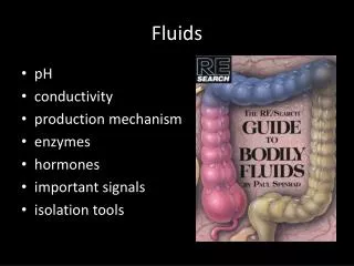
Electrolytes in body fluids • Ions form when electrolytes dissolve ad dissociate • 4 general functions • Control osmosis of water between body fluid compartments • Help maintain the acid-base balance • Carry electrical current • Serve as cofactors University of Jordan
Concentrations in body fluids • Concentration of ions typically expressed in milliequivalents per liter (mEq/liter) • Na+ or Cl- number of mEq/liter = mmol/liter • Ca2+ or HPO42- number of mEq/liter = 2 x mmol/liter • Chief difference between 2 ECF compartments (plasma and interstitial fluid) is plasma contains many more protein anions • Largely responsible for blood colloid osmotic pressure University of Jordan
ICF differs considerably from ECF • ECF most abundant cation is Na+, anion is Cl- • ICF most abundant cation is K+, anion are proteins and phosphates (HPO42-) • Na+ /K+ pumps play major role in keeping K+ high inside cells and Na+ high outside cell University of Jordan
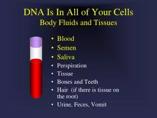
Blood • Liquid connective tissue • 3 general functions • Transportation • Gases, nutrients, hormones, waste products • Regulation • pH, body temperature, osmotic pressure • Protection • Clotting, white blood cells, proteins University of Jordan
Components of Blood • Blood plasma – water liquid extracellular matrix • 91.5% water, 8.5% solutes (primarily proteins) • Hepatocytes synthesize most plasma proteins • Albumins, fibrinogen, antibodies • Other solutes include electrolytes, nutrients, enzymes, hormones, gases and waste products • Formed elements – cells and cell fragments • Red blood cells (RBCs) • White blood cells (WBCs) • Platelets University of Jordan
I. Blood composing • Blood composing: plasma + blood cells • Hematocrit: (Packed cell volume- PCV) blood cells occupies the percentage of total blood volume. normal value male: 40-50% female: 37-48% newborn: 55%
Composition of Blood • Plasma: • Straw-colored liquid. • Consists of H20 and dissolved solutes. • Ions, metabolites, hormones, antibodies. • Na+ is the major solute of the plasma. • Plasma proteins: • Constitute 7-8 grams/100 ml of plasma. • Albumin: (MW. 68000 Dalton) • Accounts for 60-80% of plasma proteins (4.5 gm/dl) • Provides the colloid osmotic pressure needed to draw H20 from interstitial fluid to capillaries. 75% of the plasma oncotic pressure due to albumin. • Maintains blood pressure • Transport of lipid soluble hormones • Nutrition University of Jordan
Composition of the Blood (continued) • Plasma proteins (continued): • Globulins (2.5 gm/dl of plasma) (MWt. 240000 Dalton) • Maintain oncotic pressure (25% of plasma oncotic pressure) • α globulin: • Transport lipids and fat soluble vitamins. • β globulin: • Transport lipids and fat soluble vitamins. • γ globulin: • Antibodies that function in immunity. • Fibrinogen (0.5 gm/dl of plasma) • Constitutes 4% of plasma proteins. • Important clotting factor. • Converted into fibrin during the clotting process. University of Jordan
Composition of the Blood (continued) • Function of plasma protein: (1) transportation, (2) nutrition, (3) forming colloid osmotic pressure, (4) coagulation and anticoagulation, (5) pH value buffer, (6) immunity (globulin) • Serum: • Fluid from clotted blood. • Does not contain fibrinogen. • Plasma volume: • Number of regulatory mechanisms in the body maintain homeostasis of plasma volume. University of Jordan
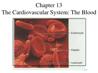
Formed Elements of Blood University of Jordan
III.Blood Cells Blood cells are erythrocyte (red blood cell, RBC), leukocyte (white blood cell, WBC) and thrombocyte (platelet, P).
Formation of Blood Cells • Negative feedback systems regulate the total number of RBCs and platelets in circulation • Abundance of WBC types based of response to invading pathogens or foreign antigens • Hemopoiesis or hemotopoiesis • Red bone marrow primary site • Pluripotent stem cells have the ability to develop into many different types of cells University of Jordan
Hematopoiesis • Undifferentiated cells gradually differentiate to become stem cells, that form blood cells. • Occurs in myeloid tissue (bone marrow of long bones) and lymphoid tissue. • 2 types of hematopoiesis: • Erythropoiesis: • Formation of RBCs. • Leukopoiesis: • Formation of WBCs. University of Jordan
Erythropoiesis • Active process. • 2.5 million RBCs are produced every second. • Primary regulator is erythropoietin. • Binds to membrane receptors of cells that will become erythroblasts. • Erythroblasts transform into normoblasts. • Normoblasts lose their nuclei to become reticulocytes. • Reticulocytes change into mature RBCs. • Stimulates cell division. • Old RBCs are destroyed in spleen and liver. • Iron recycled back to myeloid tissue to be reused in hemoglobin production. • Need iron, vitamin B12 and folic acid for synthesis. University of Jordan
Erythrocyte Physiology Shape and number of red blood cells (RBC) • Shape of RBC: like biconcave disc Its diameter is about 7~8 µm, peripheral thickness about 2.5 µm, central thickness about 1 µm and cubage about 90 µm3.
Erythrocytes • Flattened biconcave discs. • Provide increased surface area through which gas can diffuse. • Lack nuclei and mitochondria. • Half-life ~ 120 days. • Each RBC contains 280 million hemoglobin with 4 heme chains (contain iron). • Removed from circulation by phagocytic cells in liver, spleen, and bone marrow. University of Jordan
Red Blood Cells/ Erythrocytes • Contain oxygen-carrying protein hemoglobin • Production = destruction with at least 2 million new RBCs per second • Biconcave disc – increases surface area • Strong, flexible plasma membrane • Glycolipids in plasma membrane responsible for ABO and Rh blood groups • Lack nucleus and other organelles • No mitochondria – doesn’t use oxygen University of Jordan
Functions of RBC Functions of RBC • RBC can be used for transportation of O2 and CO2 in the blood. • RBC can be served as pH buffer.
Blood Cells and Platelets University of Jordan
Hemoglobin • Globin – 4 polypeptide chains • Heme in each of 4 chains • Iron ion can combine reversibly with one oxygen molecule • Also transports 23% of total carbon dioxide • Combines with amino acids of globin • Nitric oxide (NO) binds to hemoglobin • Releases NO causing vasodilation to improve blood flow and oxygen delivery University of Jordan
Shapes of RBC and Hemoglobin University of Jordan
Red Blood Cells • RBC life cycle • Live only about 120 days • Cannot synthesize new components – no nucleus • Ruptured red blood cells removed from circulation and destroyed by fixed phagocytic macrophages in spleen and liver • Breakdown products recycled • Globin’s amino acids reused • Iron reused • Non-iron heme ends as yellow pigment urobilin in urine or brown pigment stercobilin in feces University of Jordan
Formation and Destruction of RBC’s Circulation for about 120 days Circulation for about 120 days Circulation for about 120 days Circulation for about 120 days Circulation for about 120 days Circulation for about 120 days 7 7 7 7 7 7 7 3 3 3 3 3 3 3 3 3 3 3 Reused for protein synthesis Reused for protein synthesis Reused for protein synthesis Reused for protein synthesis Reused for protein synthesis Reused for protein synthesis Reused for protein synthesis Reused for protein synthesis Reused for protein synthesis Reused for protein synthesis Reused for protein synthesis Amino acids Amino acids Amino acids Amino acids Amino acids Amino acids Amino acids Amino acids Amino acids Amino acids Amino acids Transferrin Transferrin Transferrin Transferrin Transferrin Transferrin Transferrin Transferrin Fe3+ Fe3+ Fe3+ Fe3+ Fe3+ Fe3+ Fe3+ Fe3+ Globin Globin Globin Globin Globin Globin Globin Globin Globin Globin Globin Globin 6 6 6 6 6 6 6 6 4 4 4 4 4 4 4 4 4 4 5 5 5 5 5 5 5 5 5 Fe3+ Fe3+ Fe3+ Fe3+ Fe3+ Fe3+ Fe3+ Fe3+ Fe3+ Fe3+ 2 2 2 2 2 2 2 2 2 2 2 2 Fe3+ Fe3+ Fe3+ Fe3+ Fe3+ Fe3+ Fe3+ Ferritin Ferritin Ferritin Ferritin Ferritin Ferritin Ferritin Ferritin Ferritin Heme Heme Heme Heme Heme Heme Heme Heme Heme Heme Heme Heme Transferrin Transferrin Transferrin Transferrin Transferrin Transferrin Transferrin Transferrin Transferrin Transferrin + + + + + + + Globin Globin Globin Globin Globin Globin Globin Bilirubin Bilirubin Bilirubin Bilirubin 9 9 9 9 9 + + + + + + + Biliverdin Biliverdin Biliverdin Biliverdin Biliverdin Liver Liver Liver Liver Liver Liver Liver Liver Liver Vitamin B12 Vitamin B12 Vitamin B12 Vitamin B12 Vitamin B12 Vitamin B12 Vitamin B12 Bilirubin Bilirubin Bilirubin Bilirubin Bilirubin 11 11 11 1 1 1 1 1 1 1 1 1 1 1 1 1 Red blood cell death and phagocytosis Red blood cell death and phagocytosis Red blood cell death and phagocytosis Red blood cell death and phagocytosis Red blood cell death and phagocytosis Red blood cell death and phagocytosis Red blood cell death and phagocytosis Red blood cell death and phagocytosis Red blood cell death and phagocytosis Red blood cell death and phagocytosis Red blood cell death and phagocytosis Red blood cell death and phagocytosis Red blood cell death and phagocytosis + + + + + + + 10 10 10 10 Erythopoietin Erythopoietin Erythopoietin Erythopoietin Erythopoietin Erythopoietin Erythopoietin Small intestine Small intestine Small intestine Kidney Kidney 8 8 8 8 8 8 Erythropoiesis in red bone marrow Erythropoiesis in red bone marrow Erythropoiesis in red bone marrow Erythropoiesis in red bone marrow Erythropoiesis in red bone marrow Erythropoiesis in red bone marrow Bilirubin Bilirubin Bilirubin 13 13 12 12 12 Urobilin Urobilin Urobilinogen Urobilinogen Urobilinogen Macrophage in spleen, liver, or red bone marrow Macrophage in spleen, liver, or red bone marrow Macrophage in spleen, liver, or red bone marrow Macrophage in spleen, liver, or red bone marrow Macrophage in spleen, liver, or red bone marrow Macrophage in spleen, liver, or red bone marrow Macrophage in spleen, liver, or red bone marrow Macrophage in spleen, liver, or red bone marrow Macrophage in spleen, liver, or red bone marrow Macrophage in spleen, liver, or red bone marrow Macrophage in spleen, liver, or red bone marrow Macrophage in spleen, liver, or red bone marrow Macrophage in spleen, liver, or red bone marrow Key: Key: Key: Key: Key: Key: Key: Key: Key: Key: Key: Key: Key: Bacteria Bacteria Bacteria in blood in blood in blood in blood in blood in blood in blood in blood in blood in blood in blood in blood in blood Stercobilin Stercobilin Stercobilin 14 Large intestine in bile in bile in bile in bile in bile in bile in bile in bile in bile in bile in bile in bile in bile Feces Feces Feces Urine Urine University of Jordan
Starts in red bone marrow with proerythroblast Cell near the end of development ejects nucleus and becomes a reticulocyte Develop into mature RBC within 1-2 days Negative feedback balances production with destruction Controlled condition is amount of oxygen delivery to tissues Hypoxia stimulates release of erythropoietin Erythropoiesis University of Jordan
Leukocytes • Contain nuclei and mitochondria. • Move in amoeboid fashion. • Can squeeze through capillary walls (diapedesis). • Almost invisible, so named after their staining properties. • Granular leukocytes: • Help detoxify foreign substances. • Release heparin. • Agranular leukocytes: • Phagocytic. • Produce antibodies. University of Jordan
White Blood Cells/ Leukocytes • Have nuclei • Do not contain hemoglobin • Granular or agranular based on staining highlighting large conspicuous granules • Granular leukocytes • Neutrophils, eosinophils, basophils • Agranular leukocytes • Lymphocytes and monocytes University of Jordan
Types of White Blood Cells University of Jordan
Leukocyte PhysiologyClassification and numbers of Leukocyte • Number of Leukocyte (white blood cells, WBC): (4.0~11)×109/L • Classification: It is granulocyte (neutrophil, eosinophil, basophil), and agranular (monocyte and lymphocyte).
Functions of WBCs • Usually live a few days • Except for lymphocytes – live for months or years • Far less numerous than RBCs • Leukocytosis is a normal protective response to invaders, strenuous exercise, anesthesia and surgery • Leukopenia is never beneficial • General function to combat invaders by phagocytosis or immune responses University of Jordan
Many WBCs leave the bloodstream Emigration (formerly diapedesis) Roll along endothelium Stick to and then squeeze between endothelial cells Precise signals vary for different types of WBCs Emigration of WBCs University of Jordan
WBCs • Neutrophils and macrophages are active phagocytes • Attracted by chemotaxis • Neutrophils respond most quickly to tissue damage by bacteria • Uses lysozymes, strong oxidants, defensins • Monocytes take longer to arrive but arrive in larger numbers and destroy more microbes • Enlarge and differentiate into macrophages University of Jordan
WBCs • Basophila leave capillaries and release granules containing heparin, histamine and serotonin, at sites of inflammation • Intensify inflammatory reaction • Involved in hypersensitivity reactions (allergies) • Eosinophils leave capillaries and enter tissue fluid • Release histaminase, phagocytize antigen-antibody complexes and effective against certain parasitic worms University of Jordan
Physiological Characteristics and Functions of WBC • Neutrophil (60%-70%) • Another name, polymorphonuclear, PMN, 6~8 h in the vessels, diapedisis, chemotaxis and phagocytosis (using its hydrolyzed enzyme) • Function: It plays a very important role in nonspecific cellular immunity system which is against pathogenic microorganism, such as bacteria, virus, parasite, etc. • Clinic relation: Number of neutrophil greatly increase occurring in acute inflammation and earlier time of chronic inflammation. number decrease of neutrophil will result in poor resistibility and easily suffering from infection.
Physiological Characteristics and Functions of WBC • Eosinophil (2-4%) • Circadian changes: Its number is lower in the morning and higher at night. • Function: 1. It limits and modulates the effects of basophil on fast allergic reaction. 2. It is involved in immune reaction against worm with opsonization. • Clinic relation: Its number increase when person suffers from parasite infection or allergic reaction.
Physiological Characteristics and Functions of WBC • Basophil (0.5-1%) • Circulatory time: 12 hours • Basogranules contain heparin, histamine, chemotactic factors and chronic reactive material for allergic reaction. • Function: It is also involved in allergic reaction. 1. Heparin serves as lipase cobase and speeds up fatty decomposition. 2. Histamine and chronic reactive material increase permeability of capillary and contract bronchia smooth muscle, and result in allergic reaction such as measles, asthma. 3. Eosinophil chemotactic factor A released by basophil can attract eosinophil collection and modify eosinophil function.
Physiological Characteristicsand Functions of WBC • Monocyte (5-10%) • Its body is large, diameter about 15~30 µm without granule • Function: 1. It contains many nonspecific lipase and displays the powerful phagocytosis. 2. As soon as monocytes get into tissue from blood , it change name called macrophage activating monocyte- macrophage system to release many cytokins, such as colony stimulating factor (CSF), IL-1, IL-3, IL-6, TNFα, INF-α,β ,etc. 3. Cytokins induced by monocyte may modulate other cells growth. 4. Monocyte- macrophage system plays a very important role in specific immune responsive induction and regulation.
Physiological Characteristicsand Functions of WBC • Lymphocyte (20-25%) • Classification: It can be separated into T- Lymphocyte and B- Lymphocyte. • Function: 1. Lymphocytes serve as a nuclear role in immune responsive reaction. 2. T- Lymphocytes involved in cellular immunity. 3. B- Lymphocytes involved in humoral immunity. • Clinic relation: Numbers increase of lymphocytes occur in chronic inflammation and late time of infection.
Lymphocytes • Lymphocytes are the major soldiers of the immune system • B cells – destroying bacteria and inactivating their toxins • T cells – attack viruses, fungi, transplanted cells, cancer cells and some bacteria • Natural Killer (NK) cells – attack a wide variety of infectious microbes and certain tumor cells University of Jordan
Platelets (thrombocytes) • Smallest of formed elements. • Are fragments of megakaryocytes. • Lack nuclei. • Capable of amoeboid movement. • Important in blood clotting: • Constitute most of the mass of the clot. • Release serotonin to vasoconstrict and reduce blood flow to area. • Secrete growth factors: • Maintain the integrity of blood vessel wall. • Survive 5-9 days. University of Jordan
Platelets/ Thrombocytes • Myeloid stem cells develop eventually into a megakaryocyte • Splinters into 2000-3000 fragments • Each fragment enclosed in a piece of plasma membrane • Disc-shaped with many vesicles but no nucleus • Help stop blood loss by forming platelet plug • Granules contain blood clot promoting chemicals • Short life span – 5-9 days University of Jordan
Normal Value and Function of Platelet • Normal value: 100×109 ~ 300×109, range from 6%~10% • Normal changes: more number in the afternoon than in the morning, more in winter than in spring, more in the venous blood than capillary, after sport↑, pregnacy↑. • *Functions: 1. It maintains capillary endothelial cells smooth and integrated (repairing endothelium and providing nutrition). 2. It is involved in physiological hemostasis. • Platelet and clinic relation: decrease of platelet, abnormal immune reaction, will results in hemorrhage or bleeding, purpuric symptom.