Download

1 / 18
190 likes | 204 Views
WALL OF THE HEART AND CARDIAC VALVES. WALL OF THE HEART AND CARDIAC VALVES. By the end of the lecture, the student should be able to describe the microscopic structure of: 1. Wall of the heart: - Endocardium. - Myocardium.

E N D
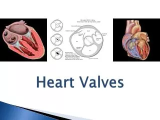
WALL OF THE HEART AND CARDIAC VALVES
WALL OF THE HEART ANDCARDIAC VALVES By the end of the lecture, the student should be able to describe the microscopic structure of: 1. Wall of the heart: - Endocardium. - Myocardium. - Epicardium. 2. Cardiac valves.
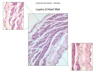
WALL OF THE HEART (A) Endocardium: 1- Endothelium 2- Subendothelial C.T. 3- Dense C.T. layer 4- Subendocardial layer (B) Myocardium (C) Epicardium: 1- Mesothelium 2- C.T. layer
ENDOCARDIUM 1- Endothelium: simple squamous epithelium. 2- Subendothelial C.T. layer 3- Dense C.T. layer 4- Subendocardial layer: • Loose C.T. layer that contains Purkinjefibers, small blood vessels & nerves. • It attaches to the endomysium of the cardiac muscle.
MYOCARDIUM • It is the middle layer • It is the most thick layer • It contains cardiac muscle cells with endomysium (loose C.T.)
CARDIAC MUSCLE • Found in the myocardium. • Striated and involuntary. • L.M. Picture of Cardiac Muscle Fibers: • Cylindrical in shape. • Intermediate in diameter between skeletal and smooth muscle fibers. • Branch and anastomose. • Covered by a thin sarcolemma. • Mononucleated. Nuclei are oval and central. • Sarcoplasm is acidophilic and shows non-clear striations (fewer myofibrils). • Divided into short segments (cells) by the intercalated discs.
Cardiac Muscle Fibers • E.M. Picture: • Few myofibrils. • Numerous mitochondria. • Less abundant SR. • T-tubules come in contact with only one cisterna of SR forming “Diads” (not triads). • Glycogen & myoglobin. • Intercalated discs: are formed of the two cell membranes of 2 successive cardiac muscle cells, connected together by junctional complexes (desmosomes and gap junctions).
EPICARDIUM(Visceral layer of pericardium) • Mesothelium:simple squamous epithelium. • Subepicardial C.T. layer: Loose C.T. contains the coronary vessels, nerves, ganglia & fat cells.
LEAFLET (CUSP) OF HEART VALVE Myocardium Endocardium Leaflet
HEART VALVES(CARDIAC VALVES) • Each leaflet (cusp) of heart valve is formed of: (1) A core of Dense irregular C.T. (2) This core is covered by: Endocardium. • The leaflets of the heart valves are normally AVASCULAR. • Blood capillaries can be found only in the base or root of the leaflet.
Wall of the Heart Practical Pictures
Endocardium Myocardium Lumen Moderator band Moderator Band