Download
1 / 2
80 likes | 288 Views
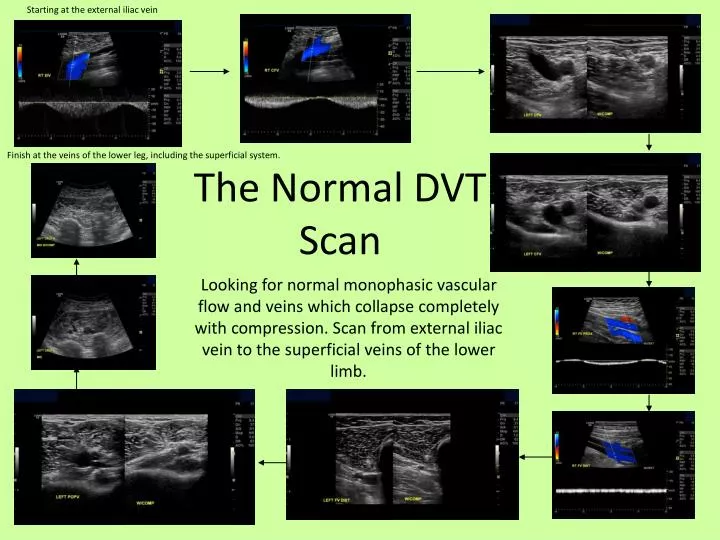
Starting at the external iliac vein. Finish at the veins of the lower leg, including the superficial system. The Normal DVT Scan.

E N D
Starting at the external iliac vein Finish at the veins of the lower leg, including the superficial system. The Normal DVT Scan Looking for normal monophasic vascular flow and veins which collapse completely with compression. Scan from external iliac vein to the superficial veins of the lower limb.
Chronic thrombus varies greatly, but may have an: * An echogenic appearance; * Dilated vessels * Retracted thrombus due to shrinkage with age. The Abnormal DVT Scan Acute thrombus can have a normal anechoic appearance to that of a normal vein. Collapse of the vein may be limited with compression, that is limited collapse will occur. Looking for changes to vascular flow and veins which do not collapse completely with compression. Vessels may appear echogenic rather than anechoic Poor compression of vessels Poor colour flow and incomplete colour fill of vessels