Download
1 / 24
260 likes | 520 Views
Vital Signs. Healthcare Science Ms. Smith. Vital Signs. Temperature, pulse, respiration, blood pressure (B/P) & oxygen saturation are the most frequent measurements taken by HCP.

E N D
Vital Signs Healthcare Science Ms. Smith
Vital Signs • Temperature, pulse, respiration, blood pressure (B/P) & oxygen saturation are the most frequent measurements taken by HCP. • Because of the importance of these measurements they are referred to as Vital Signs. They are important indicators of the body’s response to physical, environmental, and psychological stressors.
Vital Signs • VS may reveal sudden changes in a client’s condition in addition to changes that occur progressively over time. A baseline set of VS are important to identify changes in the patient’s condition. • VS are part of a routine physical assessment and are not assessed in isolation. Other factors such as physical signs & symptoms are also considered. • Important Consideration: • A client’s normal range of vital signs may differ from the standard range.
When to take vital signs • On a client’s admission • According to the physician’s order or the institution’s policy or standard of practice • When assessing the client during home health visit • Before & after a surgical or invasive diagnostic procedure • Before & after the administration of meds or therapy that affect cardiovascular, respiratory & temperature control functions. • When the client’s general physical condition changes LOC, pain • Before, after & during nursing interventions influencing vital signs • When client reports symptoms of physical distress
Body Temperature • Core temperature – temperature of the body tissues, is controlled by the hypothalamus (control center in the brain) – maintained within a narrow range. • Skin temperature rises & falls in response to environmental conditions & depends on bld flow to skin & amt. of heat lost to external environment • The body’s tissues & cells function best between the range from 36 deg C to 38 deg C • Temperature is lowest in the morning, highest during the evening.
Thermometers – 3 types • Glass mercury – mercury expands or contracts in response to heat. (just recently non mercury) • Electronic – heat sensitive probe, (reads in seconds) there is a probe for oral/axillary use (red) & a probe for rectal use (blue). There are disposable plastic cover for each use. Relies on battery power – return to charging unit after use. • Infrared Tympanic (Ear) – sensor probe shaped like an otoscope in external opening of ear canal. Ear canal must be sealed & probe sensor aimed at tympanic membrane – ret’n to charging unit after use.
Assessing Radial Pulse • Left ventricle contracts causing a wave of bld to surge through arteries – called a pulse. Felt by palpating artery lightly against underlying bone or muscle. • Carotid, brachial, radial, femoral, popliteal, posterior tibial, dorsalispedis • Assess: rate, rhythm, strength – can assess by using palpation & auscultation. • Pulse deficit – the difference between the radial pulse and the apical pulse – indicates a decrease in peripheral perfusion from some heart conditions ie. Atrial fibrillation.
Procedure for Assessing Pulses • Peripheral – place 2nd, 3rd & 4th fingers lightly on skin where an artery passes over an underlying bone. Do not use your thumb (feel pulsations of your own radial artery). Count 30 seconds X 2, if irregular – count radial for 1 min. and then apically for full minute. • Apical – beat of the heart at it’s apex or PMI (point of maximum impulse) – 5th intercostal space, midclavicular line, just below lt. nipple – listen for a full minute “Lub-Dub” • Lub – close of atrioventricular (AV) values – tricuspid & mitral valves • Dub – close of semilunar valves – aortic & pulmonic valves
Assess: rate, rhythm, strength & tension • Rate – N – 60-100, average 80 bpm • Tachycardia – greater than 100 bpm • Bradycardia – less than 60 bpm • Rhythm – the pattern of the beats (regular or irregular) • Strength or size – or amplitude, the volume of bld pushed against the wall of an artery during the ventricular contraction • weak or thready (lacks fullness) • Full, bounding (volume higher than normal) • Imperceptible (cannot be felt or heard) 0----------------- 1+ -----------------2+--------------- 3+ ----------------4+ Absent Weak NORMAL Full Bounding
Assess (cont.) • Tension – or elasticity, the compressibility of the arterial wall, is pulse obliterated by slight pressure (low tension or soft) • Stethoscope • Diaphragm – high pitched sounds, bowel, lung & heart sounds – tight seal • Bell – low pitched sounds, heart & vascular sounds, apply bell lightly (hint think of Bell with the “L” for Low)
Respirations • Assess by observing rate, rhythm & depth • Inspiration – inhalation (breathing in) • Expiration – exhalation (breathing out) • I&E is automatic & controlled by the medulla oblongata (respiratory center of brain) • Normal breathing is active & passive • Women breathe thoracically, while men & young children breathe diaphramatically ***usually • Asses after taking pulse, while still holding hand, so pt is unaware you are counting respiratons
Blood Pressure • Force exerted by the bld against vessel walls. Pressure of bld within the arteries of the body – lt. ventricle contracts – bld is forced out into the aorta to the lg arteries, smaller arteries & capillaries • Systolic- force exerted against the arterial wall as lt. ventricle contracts & pumps bld into the aorta – max. pressure exerted on vessel wall. • Diastolic – arterial pressure during ventricular relaxation, when the heart is filling, minimum pressure in arteries. • Factors affecting B/P • lower during sleep • Lower with bld loss • Position changes B/P • Anything causing vessels to dilate or constrict - medications
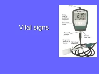
B/P (cont.) • Measured in mmHg – millimeters of mercury • Normal range • syst 110-140 dias 60-90 • Hypertensive - >160, >90 • Hypotensive <90 • Non invasive method of B/P measurement • Sphygmomanometer, stethoscope • 3 types of sphygmomanometers • Aneroid – glass enclosed circular gauge with needle that registers the B/P as it descends the calibrations on the dial. • Mercury – mercury in glass tube - more reliable – read at eye level. • Electronic – cuff with built in pressure transducer reads systolic & diastolic B/P
B/P (cont.) • Cuff – inflatable rubber bladder, tube connects to the manometer, another to the bulb, important to have correct cuff size (judge by circumference of the arm not age) • Support arm at heart level, palm turned upward - above heart causes false low reading • Cuff too wide – false low reading • Cuff too narrow – false high reading • Cuff too loose – false high reading • Listen for Korotkoff sounds – series of sounds created as bld flows through an artery after it has been occluded with a cuff then cuff pressure is gradually released. • Do not take B/P in • Arm with cast • Arm with arteriovenous (AV) • Arm on the side of a mastectomy i.e. rt mastectomy, rt arm
B/P Lower Extremity • Best position prone – if not – supine with knee slightly flexed, locate popliteal artery (back of knee). • Large cuff 1 inch above artery, same procedure as arm. Systolic pressure in legs maybe 10-40 mm hg higher • If unable to palpate a pulse – you may use a doppler stethoscope
Oxygen Saturation (Pulse Oximetry) • Non-invasive measurement of oxygen saturation • Calculates SpO2 (pulse oxygen saturation) reliable estimate of arterial oxygen saturation • Probes – finger, ear, nose, toe • Patient with PVD or Raynauds syndrome – difficult to obtain. • Normal – 90-100% • Remove nail polish • Wait until oximeter readout reaches constant value & pulse display reaches full strength • During continuous pulse oximetry monitoring – inspect skin under the probe routinely for skin integrity – rotate probe.