Download
1 / 50
540 likes | 1.4k Views
Intracellular Hormone Receptors. Steroid versus Peptide Hormones Mechanism of Action of Steroid Receptors Cellular Localization and Structure of Steroid Receptors How Steroid Receptors Initiate Transcription Role of Hormone Response Elements (HREs)

E N D
Intracellular Hormone Receptors Steroid versus Peptide Hormones Mechanism of Action of Steroid Receptors Cellular Localization and Structure of Steroid Receptors How Steroid Receptors Initiate Transcription Role of Hormone Response Elements (HREs) Interactions of Steroid Receptors with Other Pathways Regulation of Steroid Receptors
Action of Steroid Hormones versusGonadotropin Hormones Peptide HormonesSteroids Half-life in circulation short long Speed of action fast slow Duration of effect short long Location of receptor membrane inside Post-receptor regulation high low Signal amplification high low
Gene Transcription RNA mRNA R Protein Steroid Binding protein Mechanism of Action of Steroid Receptors
Tissue homogenize centrifuge Cytoplasmic fraction Nuclear fraction unoccupied occupied Cellular Localization of Steroid Receptors Early Studies (1968):
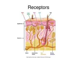
Localization of Protein by Immunocytochemistry • Incubate cell specimen with antibody which specifically detects the protein. • Wash away unbound antibody. • Use a second labeled antibody to detect the first antibody. protein
Receptor antibody Color reaction Cellular Localization of Steroid Receptors • Immunocytochemical studies indicated that all steroid receptors are nuclear.
centrifugation Cellular Localization of Steroid Receptors • Cell enucleation experiments also indicated that all steroid receptors were nuclear.
homogenize Cellular Localization of Steroid Receptors It was thought that occupied receptor had higher affinity for the nucleus than unoccupied receptor, and was thus less likely to leak out during homogenization step in earlier studies.
Low ER Expression Increased ER Expression Cellular Localization of Steroid Receptors • More recent immunocytochemistry experiments reveal cytoplasmic estrogen receptors in certain regions of the hypothalamus under certain conditions.
Cellular Localization of Steroid Receptors • It is generally thought that unoccupied steroid receptors can exist in the cytoplasm, while occupied receptors act in the nucleus on target DNA. • When bound to hormone, cytoplasmic receptors move into the nucleus.
Steroid Receptor Superfamily • Functionally Related: all are intracellular receptors which act as transcription factors, regulating target gene expression. • The Superfamily includes: glucocorticoid receptor mineralocorticoid receptor progesterone receptor (A and B) androgen receptor estrogen receptor (alpha and beta) thyroid hormone receptor vitamin D receptor retinoic acid receptor
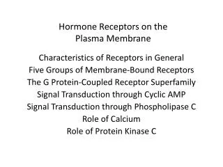
Nuclear Receptors • Proteins interact with steroids and other hormones that diffuse through the cell membrane. • Form hormone-receptor complexes that function as activators by binding to enhancers hormone response elements. • Sex hormones: estrogens and androgens; glucocorticoids, cortisol, vitamin D Ca2+ metabolism; thyroid hormone, retinoic acid developmental factors.
The majority of nuclear receptors bind to respective enhancer elements and repress transcription. - In the presence of hormone, they form R-H complexes in the nucleus and function as activators by binding to the same enhancers. - Act as repressor or enhancer, depending on the physiological signals. - thus, the response element serves as either enhancer or silencer.
Transcription Promoter Target gene Responses to hydrophobic hormones are mediated by intracellular receptors Lipophillic Hormone Plasma membrane Lipophilic hormone carried in blood Hormone binds intracellular receptor inducing receptor dimerization and activation Complex is imported into nucleus Binds to “hormone response element” to regulate gene expression Target cell Cytoplasm Intracellular receptor Translation Nuclear envelope “Hormone response element” Nucleus
2. The glucocorticoid (nuclear) receptor is found in the cytoplasm
Glucocorticoid Action 1. GR exists in an inactive form in the cytoplasm complexed with heat shock protein 90 (hsp90). 2. Glucocorticoid (G) diffuses across cell membrane and enters cytoplasm 3. G binds to GR changes conformation dissociates from hsp90 4. exposes a nuclear localization signal (stretch of aas) on GR. 5. G-GR (hormone-receptor complex, HR) enters nucleus, dimerizes with another HR.
6. HR dimer binds to enhancer/hormone-response element upstream of hormone activated gene. 7. Binding of HR dimer to enhancer activates transcription. 8. Most contain 2 zinc fingers (1) controls DNA binding, (2) controls dimerization Critical residues for discriminating between GRE and ERE lie at the base of the first finger -GRE = glucocorticoid responsive element /enhancer (sequence); ERE = estrogen
dimerization Specificity of DNA binding
Steroid Receptor Structure • This superfamily of ligand-activated transcription factors is also structurally related. • Three well conserved regions: -Hormone binding domains (HBD) in carboxyl terminus -DNA-binding domain (DBD) 5’ to ligand binding domain • A nonconserved hypervariable region, which may contribute to transcriptional activity of receptor DBD HBD hypervariable
How Are Steroid Hormone Receptors Activated? • We know that when bound to hormone, the hormone-receptor complex initiates transcription of target genes. But how? - Role of Heat Shock Proteins (HSPs) - Role of Hormone Response Elements (HREs)
Role of HSPs • Unbound receptor is associated with several HSPs. • Binding of hormone to receptor results in loss of HSPs, followed by dimerization and activation of transcription.
hsp90 H hsp70 H H R hsp70 hsp59 H H P? H H target gene HRE 5’ flanking region Steroid Receptor Action:Roles of Heat Shock Proteins and HREs What is the mechanism of steroid receptor activation? dimerize Gene Transcription
Not All Intracellular Receptors are Associated with HSPs. • HSPs bind to glucocorticoid, mineralocorticoid, androgen, progesterone, and estrogen receptors in absence of hormone. • However, receptors for thyroid hormone, retinoic acid, and vitamin D are not bound to HSPs. • This second group of receptors is bound to their hormone response element (HRE) on 5’flanking region of target genes, but are inactive until hormone binds to them.
Is Removal of HSPs Sufficient to Induce Activation of Steroid Hormone Receptors? • Removal of HSPs is not sufficient to induce transcriptional response - requires ligand. • Steroid receptors with HSP bound can still bind DNA. • Activation of steroid receptors may require a ligand-induced phosphorylation step. • Ligand-independent activation of receptors may occur if they are phosphorylated
Role of Phosphorylation in Steroid Receptor Activation • The transcriptional activity of the progesterone receptor can be stimulated by treatment with cyclic AMP. 8-bromo cyclic AMP PKA? + Phosphate DBD HBD hypervariable transcription
Role of Phosphorylation in Steroid Receptor Activation • Progesterone receptor transcriptional activity is inhibited by inhibitors of protein kinase A. Progesterone + PKA inhibitor DBD HBD hypervariable transcription
Role of Phosphorylation in Steroid Receptor Activation • Occupancy of steroid receptors by hormone is associated with increased phosphorylation of the receptor. • Thus, phosphorylation of steroid receptors appears to be an important step in receptor activation.
target gene HRE 5’ flanking region Steroid Receptors bind to Hormone Response Elements (HREs) on DNA • Following hormone binding, intracellular receptors act as transcription factors, binding to hormone response elements (HREs) on the 5’ flanking region of target genes.
Steroid Receptors bind to Hormone Response Elements (HREs) on DNA Hormone Progesterone, Androgen, Glucocorticoid, Mineralocorticoid Consensus HREs AGAACAnnnTGTTCT Estrogen AGGTCAnnnTGACCT Thyroid hormone Retinoids Vitamin D AGGTCATGACCT
H H NNN A T C G A T A T G C A T Transcription TATA EXON 1…... Palindromic Sequences Allow Binding of Receptors as Dimers 5’ -AGAACAnnnTGTTCT- 3’
Sharing of HREs by Different Steroid Receptors • Note that there are 8 classes of steroid receptors, but only 3 consensus HRE’s. Many receptors recognize the same HRE! • How is specificity achieved (how does a cell know its being stimulated by PR and not GR)? - Cell specific expression of receptors (don’t express both PR and GR in same cell. But sometimes they are in the same cell!) - Other transcriptional regulation elements (cofactors) - Formation of heterodimers versus homodimers (ie, thyroid hormone receptor with retinoic acid receptor)
Role of Cofactors in Steroid Receptor Action and Specificity • There are cofactors that interact with steroid receptors to facilitate increased transcription • Example: Cdc37 interacts with the androgen receptor, plays a role in transcriptional response. • Cdc37 affects protein folding. • Cdc37 does not interact with glucocorticoid receptor (which shares the same HRE on target DNA) Cdc37 Androgen Receptor Glucocorticoid Receptor ---AGAACAnnnTGTTCT--target gene ARE/GRE
Estrogen Receptor cDNA Transfect Cells Expression 3 H-E2 ER Evidence for HRE Specificity Mader et al., 1989 Genes with ERE= transcription Genes with GRE= no transcription
Specificity of HRE/DNA Binding Domain Interaction How do we know it’s the HRE and DNA Binding Domain that interact to give specificity of transcriptional regulation? Here’s an experiment. ERE bindingGRE bindingLigand DBD HBD WT-ER yes no estradiol HBD WT-GR no yes cortisol HBD no yesestradiol ER/GR
intron 5’-flanking region exon ERE TATA BOX How does binding of Activated Steroid Receptor to HREs Enhance Transcription? • HREs are enhancer sequences: they are orientation- and distance-independent • Binding of activated receptor to HRE may stabilize the interactions between TATA box, Transcription Factor IID, and RNA polymerase II
Ca++ Do steroids always act through classical steroid receptor mechanisms? • Some effects of steroids are observed in minutes, too quickly to be explained by regulation of transcription. • Rapid effects of steroids may involve changes in ion channels and membrane permeability, such as influencing membrane potentials in CNS neurons. • In human sperm, a membrane-bound progesterone “receptor” has been described, which may mediate the effects of P on sperm maturation. progesterone
Interactions of Steroid Receptors with Regulatory Elements of other Signaling Pathways • I previously mentioned that the transcriptional activity of the chick PR is increased by cAMP in the absence of progesterone. Protein kinase A inhibitors block this effect. • We also know that: • Glucocorticoid receptors interact with the transcription factor AP1 • Progesterone receptors can be activated by dopamine (a neurotransmitter) • Thyroid hormone receptor activity is inhibited by AP1
Macrophages Interleukin-1 Protein Kinase C Pathway Increased Expression of jun, fos (-) dimerization Glucocorticoid Receptor AP1 collagenase Molecular Mechanism through which Glucocorticoids inhibit Inflammatory Responses
Inhibition of AP-1 by GR • Glucocorticoid receptor may bind jun, decreasing the formation of AP-1 (jun/fos dimer). This results in less AP-1 to bind to the the AP-1 enhancer site on the 5’ flanking region of the collagenase gene. • However, DNA footprinting studies show that AP-1 still DOES bind the AP-1 site during GR-induced inhibition of AP-1 stimulated transcription. • GR may inhibit transcriptional activation by AP-1 once bound to the site.
Activation of Progesterone Receptor by Dopamine in absence of Progesterone • Dopamine is a common neurotransmitter in the brain. • Dopamine D1 receptor agonists mimic the effects of progesterone on female mating behavior. • The effects of dopamine can be blocked by a progesterone receptor antagonist and by antisense oligonucleotides to the progesterone receptor.
Female Lordotic Response C P D1 D1 + RU486 Activation of Progesterone Receptor by Dopamine in Absence of Progesterone -O’Malley
complementary DNA sequence AAAAA.., PR mRNA Translation Start site mRNA degradation block translation Use of Antisense Oligonucleotides to Block Gene Expression Block Gene Expression
Female Lordotic Response C P D1 D1 + antisense oligo to PR Blockade of Dopamine Activation of PR by Antisense Oligonucleotides
Estrogen Phosphorylates CREB via the MAPK Pathway – Wade & Dorsa, 2003 • Treatment of brain cells with estrogen results in rapid phosphorylation (15 min) of CREB. • This effect is blocked by an estrogen receptor antagonist (ER dependent) • This effect is dependent upon the activity of the mitogen-activated protein kinase (MAPK) pathway. • This effect is NOT dependent upon activity of protein kinase A.
E2:ER E2 MAPK pathway gene transcription (CRE) phosphorylation of CREB Estrogen Phosphorylates CREB via the MAPK Pathway – Wade & Dorsa, 2003
Regulation of Steroid Hormone Receptor Expression • In general, tissue-specific and hormone-mediated regulation of steroid hormone receptors is not as dramatic as that for peptide hormone receptors. • Autoregulation: Ligand influences expression of own receptor. • Autoregulation can occur at several levels: - transcriptional: control of gene expression - post-transcriptional: modulation of mRNA stability - post-translational: rate of receptor degradation • Examples: -Estradiol decreases uterine ER expression -Estradiol increases ER in pituitary, liver
Regulation of Steroid Hormone Receptor Expression • Heterologous regulation - Regulation by other steroids: -Estrogen up-regulates progesterone receptor in breast, uterus, hypothalamus -Progesterone down-regulates estrogen receptor -Androgen down-regulates estrogen receptor
Influences of Other Signaling Pathways • Steroid receptor expression may also be influenced by the protein kinase pathways. • Example: Activation of PKC decreases estrogen receptor mRNA stability, resulting in decreased synthesis.
Next Time… First Midterm Exam