Download
1 / 75
770 likes | 1.07k Views
Cardiopulmonary Resuscitation: Basic and Advanced Life Support.

E N D
Cardiopulmonary Resuscitation: Basic andAdvanced Life Support
Although the history of resuscitation can be traced to at least biblical times,contemporaryapproaches to cardiopulmonary resuscitation (CPR) date back to 1966, when a National Academy of Sciences National Research Council conference generated consensus standards for the performance of CPR. • Since that time, successive conferences have reviewed the practice of CPR in the light of available experimental and clinical data and have prepared revisions of previous standards. • The most recent recommendations, the “2005 American Heart Association Guidelines for Cardiopulmonary Resuscitation and Emergency Cardiovascular Care” (Guidelines 2005), represent the second internationally recognized resuscitation guidelines developed by experts from the American Heart Association (AHA) and the European Resuscitation Council; accordingly, these guidelines represent a variety of countries, cultures, and medical specialties.
Basic Life Support • BLS involves early recognition of medical emergencies, activation of an emergency response system (e.g., dialing 911 in the United States), and interventions made in response to sudden cardiac arrest (SCA), heart attack, stroke, and airway obstruction by a foreign body. • Airway, breathing, and circulation are BLS assessments readily performed without equipment. • Rescue breathing, the Heimlich maneuver, CPR, and the application-use of an automated external defibrillator (AED) are BLS interventions. • All BLS interventions are time sensitive for preventing SCA, terminating SCA, or supporting the circulation until restoration of spontaneous circulation occurs after SCA. • For those performing BLS interventions, particularly CPR, the importance of prompt initiation and expert performance of these skills cannot be overemphasized.
Cardiopulmonary Resuscitation • SCA is a complex and dynamic process. Antegrade systemic arterial blood flow continues after cardiac arrest until the pressure gradient between the aorta and right heart structures reach equilibrium. A similar process occurs during cardiac arrest with antegrade pulmonary blood flow between the pulmonary artery and the left atrium. • As the arterial-venous pressure gradients dissipate, the left heart becomes less filled, the right heart becomes more filled, and the venous capacitance vessels become increasingly distended . • When arterial and venous pressure equilibrates (approximately 5 minutes after cardiac arrest), coronary perfusion and cerebral blood flow stop.
In the absence of native cardiac function and systemic circulation, CPR is performed until return of spontaneous circulation occurs, the patient is pronounced dead, or in some special circumstances, the patient is placed on extracorporeal circulation and membrane oxygenation (ECMO). • Although CPR is far less efficient than the native circulation, when properly performed, it can provide coronary circulation and cerebral blood flow sufficient to afford full recovery in many cases if return of spontaneous circulation can be reestablished.
“Push hard and push fast” are the recommendations for chest compressions during CPR from the 2005 International Consensus conference. • Although there is insufficient evidence from human studies to identify an ideal chest compression rate, chest compressions performed at a rate of 100/min and with enough force to generate a palpable carotid or femoral pulse are considered ideal. • Chest compressions are frequently interrupted during resuscitation attempts.These interruptions have a negative impact on coronary and cerebral perfusion, as well as on return of spontaneous circulation. • Current recommendations for CPR reflect these observations by placing increased emphasis on limiting interruptions in chest compressions, even for other resuscitative measures (i.e., rescue breaths, attempts at defibrillation, tracheal intubation). • During single- and two-person CPR, Guidelines 2005 recommends compression-ventilation ratios of 30 : 2 in both scenarios with minimal interruption in chest compressions for rescue breaths.
Physiologic Considerations • Since the early 1960s, when CPR became a widespread clinical technique, it has been assumed that blood is ejected as a direct result of actual compression of the heart between the sternum and the vertebral column. This is commonly referred to as the “cardiac pump mechanism.” • Observations made with echocardiography during CPR describe a reduction in left and right ventricular volume, closure of the tricuspid and mitral valves during chest compression, and ejection of blood into the arterial system, all of which are consistent with the cardiac pump mechanism
Repeated, forceful coughing (cough CPR) can sustain consciousness during ventricular fibrillation (VF) for as long as 100 seconds when the arrhythmia (or any arrhythmia capable of rapidly compromising cardiac output) is immediately recognized and the patient can respond to verbal prompts to cough. • This observation suggests that mechanisms other than direct cardiac compression may account for forward blood flow during cardiac arrest.Vigorouscoughing produces an arterial pressure pulse associated with forward blood flow that opens the aortic valve during the generation of pressure and flow. • These findings support the proposal that increases in intrathoracic pressure generate forward blood flow,whichis commonly referred to as the “thoracic pump mechanism”.
The increasing intrathoracic pressure during chest compression equalizes intravascular pressure within the thorax. On the venous side, valve and venous collapse at the thoracic inlet limits the transmission of retrograde pressure or flow. The arterial system, which is relatively resistant to collapse, transmits pressure and flow into the extrathoracic arterial tree. • A peripheral arteriovenous pressure difference is thus established that permits blood to flow forward in the extrathoracic vascular system.
Some studies suggest that during compression or vigorous coughing the left side of the heart may act as a passive conduit for transfer of pulmonary venous blood out into the peripheral arterial circulation. During compression, blood flows from the lungs through the left ventricle toward the periphery. The pulmonary valve is closed and the mitral and aortic valves are open during periods of high intrathoracic pressure when the chest is compressed.
There is evidence that both cardiac pump and thoracic pump mechanisms exist at times during resuscitation attempts. • Echocardiographic evidence clearly demonstrates findings consistent with the cardiac pump theory. Cough CPR is effective in maintaining consciousness, at least in the early period after the onset of lethal arrhythmia, consistent with the thoracic pump theory. • Chest compressions during cardiac arrest may initially promote a cardiac pump mechanism as a result of the presence of blood in the left heart. As pressure gradients between the arterial and venous systems equilibrate during prolonged resuscitation, pulmonary blood volume increases and probably supports a thoracic pump mechanism.
Systemic, coronary, and cerebral blood flow during CPR is dependent on effective chest compressions, as well as on return of venous blood to the heart. • At chest compression rates of 80 to 100/min, the compression-relaxation ratio approaches 50 : 50. The time available for return of blood to the thorax is limited. • Venous blood returns to the thorax at very low pressure during cardiac arrest. Modest increases in intrathoracic pressure will impair return of venous blood and have a negative impact on systemic, coronary, and cerebral perfusion, in addition to reducing the chance of spontaneous circulation
Cardiac output during CPR with effective, uninterrupted chest compression is 25% to 30% of the normal spontaneous circulation. Systemic and pulmonary perfusion during CPR reflects the decreased cardiac output as demonstrated by weak carotid pulses with compression and low carbon dioxide excretion . • In cardiac arrest without a hypoxic etiology (e.g., drowning, suffocation), oxygen content in the lungs at the time of cardiac arrest is usually sufficient for maintaining an acceptable arterial oxygen content during the first several minutes of CPR. Rescue breaths are less important than initiating chest compressions immediately after the onset of SCA. Blood flow rather than arterial oxygen content is the limiting factor for delivery of oxygen to the coronary, cerebral, and systemic circulation during CPR.
Multiple modifications to standard CPR exist, as well as mechanical adjuncts to improve circulation during CPR. • A few will be reviewed here in light of current understanding.
Interposed abdominal compression (IAC) • Interposed abdominal compression (IAC) is a form of CPR in which the abdomen is compressed midway between the xiphoid process and the umbilicus during the upstroke of the chest compression phase in an attempt to sustain aortic diastolic pressure and thus improve coronary perfusion pressure, a critical determinant of successful restoration of spontaneous circulation. • Though accepted by the AHA as an satisfactory adjunct to standard CPR, this intervention is rarely used despite recognition of its benefits, minimal risk, and easy application. • When sufficient personnel trained in the technique are available, AIC-CPR can be considered a reasonable alternative to standard CPR.
Active compression-decompression CPR (ACD-CPR) • Active compression-decompression CPR (ACD-CPR) accomplishes compression and active decompression of the thorax by means of a device containing a suction header, bellows, and a compression area within the bellows. • Although these observations provided clinical support for experimental studies indicating improved hemodynamic (and perhaps ventilatory) function with ACD-CPR versus standard CPR, still lacking was evidence of benefit on patient outcome. • despite the earlier experimental and clinical evidence that ACD-CPR may confer benefit to victims of cardiac arrest by virtue of enhanced hemodynamic variables, no evidence supports the hypothesis that ACD-CPR improves outcomes after cardiac arrest in humans when compared with standard manual CPR. • The ACD-CPR device may also expose patients to harm.Currently, no ACD-CPR devices are approved by the Food and Drug Administration for sale in the United States.
The impedance threshold device (ITD) • The impedance threshold device (ITD) limits entry of air into the lungs during the recoil phase after chest compression, thereby creating greater negative intrathoracic pressure and drawing greater blood volume into the chest than with standard CPR. What results appears to be greater coronary blood flow (occurring during the diastole/chest recoil phase of chest compressions), greater cerebral blood flow, and improved hemodynamics in comparison to standard CPR. • The results of animal and clinical studies endorse the benefits of this device, which include improved resuscitation and short-term survival when compared with standard CPR. • Although this device is most effective with endotracheal tubes, if a rescuer can maintain a good seal with a facemask, the ITD can be used. • Even though long-term survival has not been documented after use of the ITD during resuscitation, if the device is available, there are no compelling reasons to not consider using this technology.
Circumferential compression of the chest with a load-distributing band (LDB) • Circumferential compression of the chest with a load-distributing band (LDB) has been described as a mechanism for generating fluctuations in intrathoracic pressure and thereby improving blood flow by exploiting the thoracic pump mechanism beyond that possible with standard CPR. • This device wraps circumferentially around the patient's torso, is closed with Velcro, and then automatically adjusts the length of the belt to fit snuggly around the patient's chest. Repetitive shortening of the belt compresses the chest and generates forward blood flow. Chest recoil occurs as the belt relaxes. As with the other devices already mentioned, the LDB has been shown in a preliminary study to increase both aortic and coronary perfusion pressure in animals, which has resulted in improved survival and neurologic outcome. • However, a recent multicenter, randomized prehospital clinical trial to evaluate the effectiveness of an LDB was halted early because of markedly reduced neurologic recovery in cardiac arrest victims on whom the LDB was used when compared with cardiac arrest victims who received standard CPR. The authors speculate that application of the device probably delayed or interrupted CPR and also noted that the time to first defibrillation in the LDB group occurred on average 2.1 minutes after the standard CPR group.
Although the devices mentioned here all demonstrate improved circulation and blood flow in comparison to standard CPR during cardiac arrest in humans, none of the devices demonstrate improved hospital discharge rates after resuscitation from cardiac arrest. • What is certain is that CPR, regardless of the mechanism of blood flow and technique, is only a temporizing procedure in need of rapid supplementation with ACLS, most critically, rapid defibrillation of the fibrillating heart.
Monitoring Performance of Cardiopulmonary Resuscitation • Until recently, palpation of the carotid or femoral pulse and observation of pupillary size were the standard and very indirect measures for assessing the apparent adequacy of CPR. Obviously, a palpable large-artery pulse indicates only the transmission of a pressure wave into the arterial tree during chest compression and provides no objective evidence of the effectiveness of cardiac output. • Initial pupillary size and changes during CPR are of some prognostic value. Pupils that are persistently contracted or initially dilated but subsequently contracting are associated with a greater likelihood of successful resuscitation and neurologic recovery than persistently dilated or subsequently dilating pupils are
Monitoring systemic arterial pressure directly, as can usually be accomplished during in-hospital CPR, is helpful in optimizing the rate and depth of chest compressions. • Aortic diastolic pressure in particular, an index of coronary perfusion pressure, should be monitored whenever possible and optimized with appropriate changes in manual compression technique and early and repeated injection of epinephrine and vasopressin before restoration of spontaneous circulation
In 1978, Kalenda described the use of capnographyas a guide to the effectiveness of external chest compressions. He demonstrated the value of monitoring Petco2in three patients in cardiac arrest and confirmed changes in Petco2 with restoration of spontaneous circulation. Kalenda proposed that when ventilation is constant, as during controlled mechanical ventilation, “the expired CO2 is a precise and continuous mirror of lung perfusion and hence of cardiac output.”
Evidence is increasing that Petco2 measurements obtained during cardiac arrest and CPR may have predictive value relative to the likelihood that spontaneous circulation will be restored. • Although more such data are needed to quantitate the predictive power of Petco2, it seems certain that Petco2 measurements during cardiac arrest and CPR will become an objective index to predict the likelihood that persistent resuscitative effort will result in restoration of spontaneous circulation.
Petco2 reflects pulmonary blood flow and therefore cardiac output. • Monitoring of both systemic arterial pressure by arterial catheter and Petco2 with controlled ventilation should provide optimal hemodynamic assessment of the adequacy of the resuscitative effort and response to interventions such as changes in depth, rate, and location of manual chest compressions, as well as response to drugs such as epinephrine and vasopressin.
Compelling evidence confirms major deficiencies in the provision of CPR, particularly by trained health care professionals, in both out-of-hospital and in-hospital settings. • Long pauses without chest compressions and compressions that are suboptimal in both rate and depth are frequent in both settings and result in decreased blood flow. Animal studies have shown that inadequate blood flow from poorly performed CPR during cardiac arrest has a negative impact on both restoration of spontaneous circulation and survival. • Clinical evidence also suggests that excessive ventilation occurs during resuscitation attempts and likewise has a negative impact on resuscitation outcomes. • In light of these observations, the major focus in Guidelines 2005 simplified CPR for the purpose of prolonging uninterrupted chest compression.
Airway Control and Ventilation • Ventilation is critical for restoration of spontaneous circulation and organ preservation during cardiac arrest. The AHA ACLS training program has effectively conveyed this treatment priority. • The techniques used for providing ventilation are obviously dependent on the clinical situation. The head tilt–chin lift maneuver is recommended for initial airway control. • The epiglottis rather than the tongue is the major cause of upper airway obstruction in unconscious humans.Because of its ligamentous attachments to the hyoid bone, the epiglottis can be lifted by manual maneuvers that displace the hyoid bone anteriorly. These observations provide anatomic confirmation of the efficacy of the head tilt–chin lift technique for opening an obstructed airway. The head tilt–jaw thrust maneuver accomplishes the same purpose in restoring airway patency.[
When rescue breathing is indicated for a nontracheallyintubated cardiac arrest victim, two 1-second breaths are delivered after the 30th compression during both one- and two-person CPR. • Rescue breaths should provide only enough force and volume to cause chest rise. Excessive ventilation force or tidal volumes run the risk of overcoming esophageal opening pressure and thereby contributing to gastric inflation and its consequences. • Once a tracheal tube is in place, ventilation can occur at a rate of 8 to 10 breaths per minute independent of chest compressions. • Ventilation should minimally disrupt chest compressions.
During the first few minutes after the onset of cardiac arrest, chest compressions are more important than rescue breathing (provided that the cardiac arrest is not secondary to asphyxiation, as with drowning or suffocation). • Delivery of oxygen to tissues with CPR is limited more by blood flow and low cardiac output than by arterial content.The low cardiac output associated with CPR results in low oxygen uptake from the lungs, which in turn reduces the need to ventilate the patient during this low-flow state. • In view of this information, uninterrupted chest compressions must be given the highest priority early in resuscitation efforts.
Endotracheal intubation is the usual and expected standard of airway control in the critical care setting.Alternative airways that may be useful in gaining rapid control of the airway and ventilation while reducing the risk of pulmonary aspiration of gastric contents in situations in which tracheal intubation is not possible include the laryngeal mask airway (LMA) and the esophageal-tracheal Combitube. These devices are classified in Guidelines 2005 as acceptable and possibly helpful, especially when the rescuer is inexperienced in placing tracheal tubes. • Although they may be helpful as temporizing devices, endotracheal intubation remains the optimal technique for controlling the airway and ventilating the lungs during CPR. Regardless of the device used, once an advanced airway is inserted and placement confirmed, ventilation can be provided at a rate of 8 to 10/min without interruption of chest compressions.
Confirmation of proper placement of an endotracheal tube • Confirmation of proper placement of an endotracheal tube can be difficult in a patient who has undergone cardiac arrest. Observation of the rise and fall of the thorax and auscultation of lung fields in this situation can be misleading. Likewise, because of the very low pulmonary blood flow during CPR, Petco2 detection devices may not readily distinguish tracheal from esophageal intubation.
For these reasons, esophageal detector devices based on the description by Weehave been introduced and advocated for use in emergency situations such as cardiac arrest. Both a syringe and a self-inflating bulb have been used. • The efficacy of these devices in distinguishing esophageal from tracheal intubation is based on the principle that the trachea remains patent during aspiration of air whereas the esophagus collapses because of its fibromuscular structure. • The effectiveness of the self-inflating bulb in distinguishing esophageal from tracheal tube position and in confirming proper position of the esophageal-tracheal tube has been documented.
esophageal detector device • In an emergency patient population, an esophageal detector device as described by Wee was observed to be more accurate than detection of Petco2 because of its greater accuracy in patients in cardiorespiratory arrest .
False-negative results with the self-inflating bulb occur more frequently in emergency intubations than in anesthetized patients undergoing elective procedures. Causes include partial tube obstruction with secretions, atelectasis, bronchospasm, and endobronchial intubation.A high incidence of false-negative results was observed in morbidly obese patients.In these patients, reduced functional reserve capacity and large-airway collapse secondary to invagination of the membranous posterior tracheal wall after the application of subatmospheric pressure with the self-inflating bulb were identified as the cause of the false-negative results. • With these limitations in mind, the self-inflating bulb or syringe-type esophageal detector is useful in emergency situations such as cardiac arrest, particularly when used in combination with Petco2 detection.
LMA • Despite extensive experience with the LMA in fasted patients undergoing general anesthesia, its role during CPR remains somewhat controversial.In anesthetized patients, positive-pressure ventilation with the LMA is safe and effective, but concern was expressed that gastric inflation could be a problem in the presence of increased inflation pressure.This problem, of course, is common in patients who sustain cardiorespiratory arrest because they typically have a full stomach and frequently require high inflation pressure during ventilation. • The LMA has been used successfully in arrested patients who have no evidence of regurgitation or aspiration. • In patients in whom endotracheal intubation is not possible, the LMA is more secure than a facemask and offers an alternative to control the airway and ventilate the patient. • Importantly, placement of an LMA by an inexperienced provider cannot result in unrecognized esophageal intubation in a patient requiring emergency airway management.
The esophageal-tracheal Combitube is an acceptable alternative airway device for use in cardiac arrest. The Combitube is a double-lumen device with proximal pharyngeal and distal inflatable cuffs that is introduced blindly into the airway. One lumen of the device has a closed distal lumen and ventilation holes at the level of the hypopharynx. The second lumen is open ended with a balloon cuff at its distal end. Both lumens of the airway can accommodate ventilation. Once inserted and followed by inflation of the pharyngeal and distal cuffs, confirmation of the proper ventilation port is mandatory. Auscultation and Petco2 detection must demonstrate that tracheal rather than esophageal ventilation has been achieved. • When the esophageal-tracheal Combitube is properly placed, the airway is isolated and the risk for aspiration of gastric contents is reduced.
If these devices and techniques are unsuccessful in securing an airway, immediate cricothyroidotomy may be necessary. A 12-, 13- or 14-gauge catheter-over-needle device can be inserted quickly into the trachea through the cricothyroid membrane. Equipment permitting transtracheal jet ventilation from a 50-psi oxygen source through such a catheter should be available in the operating room and ICU.
Increasingly, airway management and ventilation conducted in the setting of resuscitation has come under scrutiny.Clinical studies support the concept that circulation, not ventilation, has the greatest impact on survivability after cardiac arrest.Yet chest compressions are frequently interrupted for prolonged periods to allow either ventilation or placement of an advanced airway in both the in-hospital and prehospital environments. • Ventilation during resuscitation, with or without the advanced airway in place, is often excessive and associated with increased intrathoracic pressure, lower coronary perfusion pressure, impaired venous return, and ultimately, decreased survival. • The team leader must be attentive to the rate and vigor of ventilation performed during resuscitation attempts, in addition to other components of therapy.
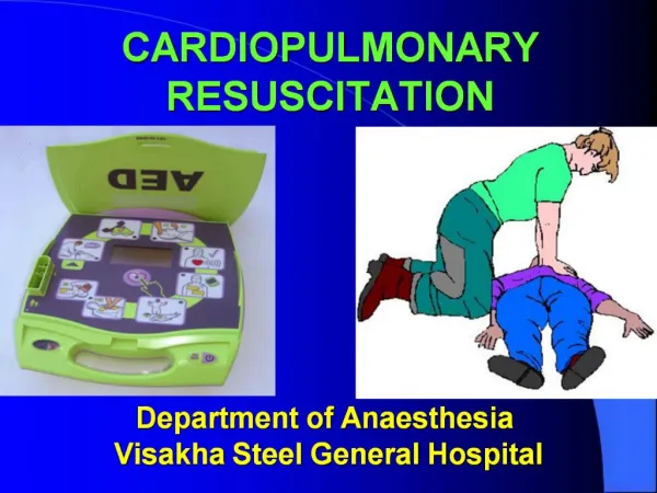
Automated External Defibrillators and Manual Defibrillation • The most frequent cardiac rhythm responsible for witnessed cardiac arrest in adults is VF. CPR prolongs the duration of VF but cannot convert the arrhythmia to an organized rhythm. Successful termination of this arrhythmia requires prompt electrical defibrillation, not medications. The most recent AHA guidelines include early defibrillation with an AED as part of BLS training and the concept of public-access defibrillation, which endorses the policy of making defibrillation available to victims of cardiac arrest through nonconventional providers (e.g., police, security guards, and others).The AED, when applied to a patient, is capable of analyzing cardiac rhythm, detecting VF and rapid ventricular tachycardia (VT), and then delivering a defibrillatory shock. A trained rescuer's role is to apply the defibrillator pads to the patient's chest, activate the AED, and if the device indicates that a shock is indicated, deliver the shock by pushing a button when prompted to do so by the AED.
Previous guidelines addressing AED use in cardiac arrest recommended administering up to three consecutive uninterrupted shocks if needed to terminate VF. • Chest compressions were not performed during the shock sequence. • Current experimental and clinical evidence suggests that success in defibrillation and survival are negatively influenced by frequent or prolonged interruptions in chest compressions during cardiac arrest.
In light of this most recent information, Guidelines 2005 recommends a single shock from an AED followed immediately by a 2-minute period of CPR, the equivalent of five cycles of 30 : 2 chest compression-ventilation, before reanalysis of the cardiac rhythm. • Chest compressions are continued until the AED is charged and ready to deliver the shock. • In most instances, the heart is at least transiently “stunned” by defibrillation shocks and is benefited by a period of coronary blood flow with chest compressions. • For the exceptional patient with invasive monitoring present at the time of cardiac arrest, if an organized cardiac rhythm with a perfusing arterial waveform follows defibrillation, chest compressions may not be indicated.
VF is a highly metabolically active state of the myocardium. In the presence of VF, myocardial stores of oxygen and metabolic substrates are depleted rapidly. Chest compressions deliver oxygen and energy substrates to the myocardium, thus making defibrillation more likely. • Experimental animal and prehospital evidence from Seattleand Oslodemonstrate increased defibrillatory success when CPR is provided before defibrillation, particularly if the cardiac arrest was unwitnessed or if the time between cardiac arrest and arrival of rescuers exceeds 4 minutes.
Guidelines 2005 recommends 2 minutes of CPR (five cycles of 30 : 2 compression-ventilation) before the first rhythm analysis of the cardiac arrest in the setting of unwitnessed cardiac arrest or delays in initiation of CPR. • In settings in which defibrillation technology (manual defibrillator or AED) is immediately available (i.e., <3 minutes), rescuers should begin CPR and then defibrillate as soon as possible.
If a monophasic defibrillator is available, defibrillation energy should begin at high energy (300 to 360 J). Prospective human clinical studies have failed to identify the optimal biphasic energy levels for first or subsequent defibrillation attempts.Although first-shock energies specific to the defibrillation waveform have been recommended (150 to 200 J with a biphasic truncated exponential waveform, 120 J with a rectilinear biphasic waveform), a rescuer encountering an unfamiliar device should choose an energy setting of 200 J for the first shock and may increase the energy as needed for subsequent shocks when indicated. • when biphasic waveform defibrillation is used, the body weight of the patient does not influence the energy delivered because the waveforms compensate for transthoracic impedance to allow uniform delivery of energy and thus obviate the need to vary from the recommended defibrillation energy. • Time to the first shock remains the most important factor influencing successful resuscitation
Advanced Cardiac Life Support • It must be emphasized that CPR almost invariably necessitates rapid interventional follow-up care with ACLS procedures. Anesthesiologists should be capable of rendering such definitive follow-up intervention, whether in the operating room, ICU, emergency department, delivery room, or hospital ward. • The somewhat reassuring observation that intraoperative cardiac arrests are rare (1.1/10,000 and 1.4/10,000 in the two studies) does not dismiss the need for anesthesiologists to be thoroughly acquainted with ACLS equipment and interventions because when these methods are needed, they must be executed skillfully and decisively. • Failure to intervene rapidly with ACLS pharmacologic therapy was identified as a major cause of poor outcome in other reports of intraoperative cardiac arrest.
In a study using a computer program that simulates critical patient incidents such as cardiac arrest, it was observed that only 30% of participants, who consisted of anesthesiology residents, faculty, and private practitioners, managed a simulated cardiac arrest in accordance with the AHA ACLS guidelines. • Timing since the last ACLS training was noted to be an important predictor of proper management of simulated cardiac arrest. • some form of periodic training and retraining in ACLS is necessary for maintaining the level of knowledge and skills essential for management of cardiorespiratory arrest in accord with contemporary principles as incorporated in the ACLS training program.