Download
1 / 19
270 likes | 669 Views
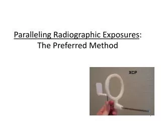
The Paralleling Technique- Part 1. Reference Reading: Chapter 17; pp. 156-158 (up to “step-by-step procedures”). The Paralleling Technique. Before the dental radiographer can competently perform the paralleling technique, he/she must have a thorough understanding of Terminology Principles

E N D
The Paralleling Technique- Part 1 Reference Reading: Chapter 17; pp. 156-158 (up to “step-by-step procedures”)
The Paralleling Technique • Before the dental radiographer can competently perform the paralleling technique, he/she must have a thorough understanding of • Terminology • Principles • Basic rules that govern this technique
Terminology • Parallel: moving or lying in the same plane, always separated by the same distance and not intersecting
Terminology • Intersecting: to cut across or through
Terminology • Perpendicular: intersecting at, or forming a right angle
Terminology • Right Angle: an angle of 90 degrees formed by 2 lines perpendicular to one another
Terminology • Long Axis of the tooth: an imaginary line that divides the tooth longitudinally (from tip of crown to tip of root), into two equal halves
Terminology • Central Ray: the center portion of the primary beam of radiation.
Principles of the Paralleling Technique OPEN TO PAGE 156 OF YOUR TEXTS
Basic Principles • The film is placed in the mouth PARALLEL to the long axis of the tooth being radiographed.
Basic Principles • The central ray is directed perpendicular to both film and tooth
Basic Principles • A beam alignment device must be used to hold the film parallel with the tooth. • The patient cannot hold the film in this manner.
Basic Principles-increasedobject-receptor distance • On maxillary arch, the film must be placed toward the middle of the oral cavity in order to achieve parallelism. • This can result in increased image magnification.
Basic Principles- Increased target-receptor distance • To compensate for the image magnification, we use a “long cone” in the paralleling technique. • Sometimes referred to as “The Long Cone Technique”
Basic Principles • Using a long cone ensures that only the most parallel rays at the center of the x-ray beam will be directed at the tooth and film.
Now…..to Review! • Film must be placed parallel to the tooth • The central ray must be directed perpendicular to both film and tooth.
REVIEW • Film will be placed at mid-palate on the maxillary shots. (increased object-receptor distance) • This increased DISTANCE between the film and the tooth can result in MAGNIFICATION.
Review • To COMPENSATE for this magnification, we use a LONG CONE (16 inches). (increased target-receptor distance) • This ensures that only the rays near the center of the x-ray beam form the image.