Download
1 / 47
470 likes | 602 Views
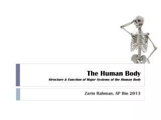
The Human Body: An Orientation Ch . 1a. Overview of Anatomy and Physiology. Anatomy: Physiology:. Principle of Complementarity. Anatomy and physiology are inseparable. Levels of Structural Organization. Chemical: Cellular: Tissue: Organ: Organ system: Organismal:. Organelle. Atoms.

E N D
Overview of Anatomy and Physiology • Anatomy: • Physiology:
Principle of Complementarity • Anatomy and physiology are inseparable.
Levels of Structural Organization • Chemical: • Cellular: • Tissue: • Organ: • Organ system: • Organismal:
Organelle Atoms Molecule Smooth muscle cell Cellular level 2 Chemical level 1 Smooth muscle tissue Cardiovascularsystem Tissue levelTissues consist of similartypes of cells. 3 Heart Bloodvessels Blood vessel (organ) Smooth muscle tissue Connective tissue Epithelialtissue Organ levelOrgans are made up of different typesof tissues. 4 Organismal levelThe human organism is made upof many organ systems. Organ system levelOrgan systems consist of differentorgans that work together closely. 6 5 Figure 1.1
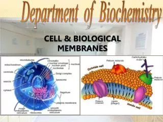
Hair Nails Skin • Integumentary System – Maintains boundaries: • Cellular Membranes • Skin Figure 1.3a
Bones Joint • Skeletal System – • Movement • Protects and supports body organs • Attachment site for muscles • Mineral storage • Blood cell formation Figure 1.3b
Skeletal muscles • Muscular System • Locomotion, facial expression • Maintains posture • Produces heat. Figure 1.3c
Brain Nerves Spinal cord • Nervous System • Fast-acting control system of the body • Responds to internal & external changes by activating • appropriate muscles and glands. Figure 1.3d
Pineal gland Pituitary gland Thyroid gland Thymus Adrenal gland Pancreas Testis Ovary • Endocrine System • Glands secrete hormones that regulate • processes various processes Figure 1.3e
Heart Blood vessels • Cardiovascular System • Blood vessels transport blood, • whichcarries oxygen, carbon • dioxide,nutrients, wastes, etc. • The heart pumpsblood. Figure 1.3f
Red bone marrow Thymus Lymphatic vessels Thoracic duct Spleen Lymph nodes • (Lymphatic System/Immunity • Picks up fluid leaked from blood vessels • and returns it to blood • Disposes of debris in lymphatic stream • Houses WBC’s involved in immunity. Figure 1.3g
Nasal cavity Pharynx Bronchus Larynx Trachea Lung • Respiratory System • Keeps blood constantly supplied with oxygen and removes carbon dioxide. Figure 1.3h
Oral cavity Esophagus Liver Stomach Small intestine Large intestine Rectum Anus • Digestive System • Breaks down food into absorbable units that enter blood • for distribution to body cells. Indigestible foodstuffs are eliminated as feces. Figure 1.3i
Kidney Ureter Urinary bladder Urethra • Urinary System • Eliminates nitrogenous wastes • Regulates water, electrolyte and • acid-base balance of the blood. Figure 1.3j
Mammary glands (in breasts) Prostate gland Ovary Penis Ductus deferens Testis Uterine tube Scrotum Uterus Vagina (Female Reproductive System Male Reproductive System Overall function is production of offspring. Figure 1.3k-l
Digestive system Takes in nutrients, breaks them down, and eliminates unabsorbed matter (feces) Respiratory system Takes in oxygen and eliminates carbon dioxide Food O2 CO2 Cardiovascular system Via the blood, distributes oxygen and nutrients to all body cells and delivers wastes and carbon dioxide to disposal organs Blood CO2 O2 Urinary system Eliminates nitrogenous wastes and excess ions Heart Nutrients Interstitial fluid Nutrients and wastes pass between blood and cells via the interstitial fluid Integumentary system Protects the body as a whole from the external environment Feces Urine Figure 1.3
Survival Needs • Nutrients • Oxygen • Water • Normal body temperature • Appropriate atmospheric pressure
Homeostasis • Definition: • A dynamic state of equilibrium
Components of a Homeostatic Control Mechanism • Receptor (sensor) • Control center • Effector
4 Output:Information sent alongefferent pathway toeffector. 3 Input: Informationsent along afferentpathway to controlcenter. ControlCenter Afferentpathway Efferentpathway 2 Receptor Effector 5 Receptordetectschange. Responseof effectorfeeds backto reducethe effect ofstimulusand returnsvariable tohomeostaticlevel. 1 IMBALANCE Stimulusproduceschange invariable. BALANCE IMBALANCE Figure 1.4
Negative Feedback • The response reduces or shuts off the original stimulus • Example: • Regulation of body temperature
Control Center (thermoregulatory center in brain) Information sent along the afferent pathway to control center Information sent along the efferent pathway to effectors Efferent pathway Afferent pathway Receptors Temperature-sensitive cells in skin and brain Effectors Sweat glands Sweat glands activated Response Evaporation of sweat Body temperature falls; stimulus ends Stimulus Body temperature rises BALANCE Stimulus Body temperature falls Response Body temperature rises; stimulus ends Receptors Temperature-sensitive cells in skin and brain Effectors Skeletal muscles Afferent pathway Efferent pathway Shivering begins Information sent along the efferent pathway to effectors Information sent along the afferent pathway to control center Control Center (thermoregulatory center in brain) Figure 1.5
Positive Feedback • The response enhances or exaggerates the original stimulus • Rare in biological systems • Example: • Enhancement of labor contractions by oxytocin
1 Break or tearoccurs in bloodvessel wall. Positive feedbackcycle is initiated. 3 2 Releasedchemicalsattract moreplatelets. Plateletsadhere to siteand releasechemicals. Positivefeedbackloop Feedback cycle endswhen plug is formed. 4 Platelet plugforms. Figure 1.6
Anatomical Position • Standard anatomical body position: • Body erect • Feet slightly apart • Palms facing forward
Regional Terms • Regional Terms: designate specific areas/regions of the body.
Upper limb Acromial Brachial (arm) Orbital Antecubital Nasal Antebrachial (forearm) Oral Carpal (wrist) Cervical Thoracic Axillary Digital Sternal Abdominal Lower limb Umbilical Coxal (hip) Pelvic Femoral (thigh) Inguinal Patellar Crural (leg) Fibular Pubic Tarsal (ankle) Thorax Abdomen Back (Dorsum) (a) Anterior/Ventral Figure 1.5
Upper limb Cephalic Acromial Occipital (back of head) Brachial (arm) Olecranal Cervical Back (dorsal) Scapular Vertebral Digital Lumbar Sacral Femoral (thigh) Gluteal Popliteal Sural (calf) Fibular Calcaneal Plantar (b) Posterior/Dorsal Figure 1.5
Body Planes and Sections • Sagittal plane • Midsagittal (median) plane • Parasagittal plane • Frontal (coronal) plane • Transverse (horizontal) plane
Frontal plane Median (midsagittal) plane Transverse plane (a) Frontal section (through torso) (b) Transverse section (through torso, inferior view) (c) Median section (midsagittal) Pancreas Aorta Spleen Liver Spinal cord Intestines Rectum Spleen Left and right lungs Liver Heart Body wall Vertebral column Stomach Arm Subcutaneous fat layer Figure 1.6
Body Cavities • Two Large Cavities: • Dorsal cavity • Two subdivisions: • Cranial cavity • Vertebral cavity
Body Cavities • Ventral cavity • Two subdivisions (separated by diaphragm):
Cranial cavity Dorsal body cavity Ventral body cavity Cranial cavity Vertebral cavity Thoracic Cavity Dorsal body cavity Vertebral cavity Ventral body cavity (thoracic and abdominopelvic cavities) Diaphragm Abdominal cavity (contains digestive viscera) Abdomino- pelvic cavity Pelvic cavity (contains urinary bladder, reproductive organs, and rectum) (a) Lateral view (b) Anterior view Figure 1.7
Ventral Body Cavities • Thoracic cavity subdivisions: • Two pleural cavities • Mediastinum • Pericardial cavity
Ventral Body Cavities • Abdominopelvic cavity subdivisions: • Abdominal cavity • Pelvic cavity
Cranial cavity Dorsal body cavity Ventral body cavity Cranial cavity Vertebral cavity Thoracic cavity Dorsal body cavity Vertebral cavity Ventral body cavity Diaphragm Abdominal cavity Abdomino- pelvic cavity Pelvic cavity (a) Lateral view (b) Anterior view Figure 1.7
Abdominopelvic Regions • Nine divisions used primarily by anatomists
Diaphragm Liver Right hypochondriac region Left hypochondriac region Epigastric region Stomach Gallbladder Transverse colon of large intestine Ascending colon of large intestine Right lumbar region Left lumbar region Umbilical region Descending colon of large intestine Small intestine Cecum Initial part of sigmoid colon Right iliac (inguinal) region Hypogastric (pubic) region Left iliac (inguinal) region Appendix Urinary bladder (a) Nine regions delineated by four planes (b) Anterior view of the nine regions showing the superficial organs Figure 1.12
Abdominopelvic Quadrants • Divisions used primarily by medical personnel
Right upper quadrant (RUQ) Left upper quadrant (LUQ) Right lower quadrant (RLQ) Left lower quadrant (LLQ) Figure 1.11