Download

1 / 47
530 likes | 756 Views
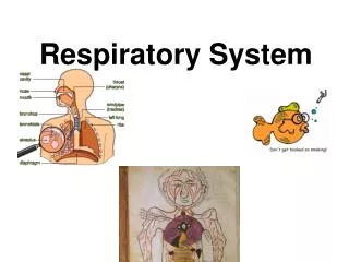
Respiratory Disorders: Pleural & Thoracic Injury. by Charlotte Cooper RN, MSN, CNS modified by Kelle Howard RN, MSN. Thoracic Cavity.

E N D
Respiratory Disorders: Pleural & Thoracic Injury by Charlotte Cooper RN, MSN, CNS modified by Kelle Howard RN, MSN
Thoracic Cavity http://www.google.com/imgres?imgurl=http://www.tcnj.edu/~mckinney/body.jpg&imgrefurl=http://www.tcnj.edu/~mckinney/breathing.htm&h=480&w=460&sz=60&tbnid=7I0hIqYYrrrEDM:&tbnh=129&tbnw=124&prev=/images%3Fq%3Dthoracic%2Bcavity&usg=__64_qfRrbnmkZHJsDtcpsNyD-QUk=&ei=HjqESrfvF4eosgPnloGqBw&sa=X&oi=image_result&resnum=4&ct=image
Normal Anatomy • Thoracic cavity • Chest wall • Pleural space • Fluid
Terminolgy • Pleura • the thin serous membrane around the lungs and inner walls of the chest (2 layers) • Pleural space • thin space between the 2 layers of pleura • Pleural cavity • body cavity that surrounds the lungs • Pleural Fluid • pleura that lines the inner chest wall and covers the diaphragm
Pleural Fluid • pH 7.6 – 7.64 • 1-2g/dL protein • Less than 1000 WBC per cubic millimeter • Glucose level similar to plasma • LDH less than 50% that of plasma • Na, K+, & Ca levels similar to that of interstitial fluid
Viceral pleura – • Covers surface of the lung • Cannot be disected away from the lung • Parietal pleura- • Lines the wall of the chest and covers the diaphragm http://www.themesotheliomalibrary.com/pleural-effusions.JPG
Chest Trauma & Thoracic Injury • 20-25% of trauma victims with chest trauma die • 45% of trauma victims have some type of chest trauma • BEWARE: External injury may appear minor
Categories for Traumatic Injuries • Blunt trauma • Penetrating trauma
What needs to be done? • Client comes to ED following a MVA • Assessment • Respiratory • Cardiovascular • Surface findings • Interventions • Monitoring • Diagnostic Test
Respiratory Disorders: Pleural and Thoracic Injury • Pleural Effusion • A collection of excess fluid in the pleural space • Classification • Transudative aka: hydorthoraces • Exudative
hydrostatic pressure or oncotic pressure • Pathophysiology of Pleural Effusion capillary permeability Formation of excess fluid= Transudate Formation of fluid & cells= Exudate
Empyema • What is it? • What causes it? • How do we treat it?
What are some causes of: • Transudative • Exudative
Clinical Manifestations: Pleural Effusion • Dyspnea • Pleurisy • Decreased breath sounds • Decreased chest wall movement • Dullness on percussion
How do we diagnosis pleural effusions?
Pleural Effusion -- Diagnositcs • ____________ • ____________ • ____________ • ____________
How do we know what type of pleural effusion it is?
Interventions: Pleural Effusion • Thoracentesis Diagnostic vs. Therapeutic
Interventions: Pleural Effusion • Treat underlying condition – • CHF/Renal failure • Pneumonia • Liver Disease • Lupus/RA • Malignancy • Pleurodesis • Chest tube insertion • Allow to resolve
Complications of Pleural Effusion • Trapped Lung • Recurrent effusions • Pneumothorax
PNEUMOTHORAX • 3 types • Closed • Open • Iatrogenic
ww http://images.google.com/imgres?imgurl=http://graphics8.nytimes.com/images/2007/08/01/health/adam/15210.jpg&imgrefurl=http://www.nytimes.com/slideshow/2007/08/01/health/100150Pneumothoraxseries_4.html&usg=__VZn79dHtqdr7izJf1jBM0r5R4ig=&h=320&w=400&sz=44&hl=en&start=3&sig2=06HaoI7v1pH1SPxnpU_4Vg&um=1&tbnid=l0LTfAdhVxUVSM:&tbnh=99&tbnw=124&prev=/images%3Fq%3Dblebs%2Bon%2Blungs%2Bcausing%2Bpneumothorax%26hl%3Den%26rlz%3D1T4DMUS_enUS282US282%26sa%3DN%26um%3D1&ei=lGWJSvXaGIawtAOVxtidBw
Clinical Manifestations: Pneumothorax • Respiratory • Cardiac
Tension Pneumothorax • Air/blood/fluid rapidly entering the pleural space • Lung collapses • Emergency situation
Pathophysiology: Tension Pneumo • Increase in intrapleural pressure • Compression of lung • Compresses against trachea, heart, aorta, esophagus • Ventilation and cardiac output greatly compromised
Clinical Manifestations: Tension Pneumo • Severe dyspnea • Tracheal deviation • Decreased cardiac output • Distended neck veins • Increased respiratory rate • Increased heart rate • Decreased blood pressure • Shock
Treatment Tension Pneumo • Emergency --- quick intervention • Needle decompression • Chest tube placement
Other Types • Hemothorax • Chylothorax
Intervention: Pneumothorax • High Fowlers position • Oxygen as ordered • Rest to decrease oxygen demand • ***Chest tube insertion • Pleurodesis • Surgery ?
Trauma of the Chest/Lung • What is involved • Chest wall • Lungs • Heart and great vessels • Esophagus • Airway obstruction • Pneumothorax • Flail chest
Clinical Manifestations: Rib Fractures • Ribs 5-10 most commonly fractured • Pain • Splinting & Rapid, shallow respirations • Decreased breath sounds • Crepitus • Signs/symptoms of pneumothorax
Treatment: Rib Fractures • Reduce or minimize pain • Do we wrap or bind the chest? • Do we use opiods? • Goal?
Pathophysiology: Flail Chest • 2 or more ribs fractured • 2 or more separate places • Unstable / free floating chest • Usually involves anterior or lateral fx • Paradoxical respirations
Clinical Manifestations: Flail Chest • Dyspnea with rapid, shallow inspiration • Pain • Palpable crepitus • Decreased breath sounds • Unequal chest expansion • Tachycardia
Interventions: Flail Chest • Oxygen as ordered • Elevate HOB • Analgesia • Suction • Splint affected side • *Intubation • *Mechanical ventilation
Pathophysiology: Pulmonary Contusion • Abrupt chest compression then rapid decompression • Intra-alveolar hemorrhage • Interstitial/bronchial edema • Decrease surfactant production • Increase pulmonary vascular resistance • Decrease blood flow
Clinical Manifestation: Pulmonary Contusion • Increased SOB • Restlessness • Anxiety • Chest pain • Copious sputum • Increased respiratory • Increased heart rate • Dyspnea • Cyanosis
Intervention: Pulmonary Contusion • Intubation • Mechanical ventilation • Bronchoscopy • Fluids • Volume expanders • Pulmonary artery pressure monitoring