Download
1 / 34
340 likes | 514 Views
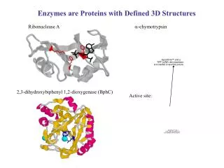
Proteins and Enzymes BIOL 239 & BMSC 209 Enzyme Function Paul Teesdale-Spittle KK712, Ext 6094, e-mail: paul.teesdale-spittle@vuw.ac.nz. The catalytic power and specificity of an enzyme is a product of the protein structure . structural complimentarity with the substrate

E N D
Proteins and Enzymes BIOL 239 & BMSC 209 Enzyme Function Paul Teesdale-Spittle KK712, Ext 6094, e-mail: paul.teesdale-spittle@vuw.ac.nz
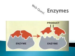
The catalytic power and specificity of an enzyme is a product of the protein structure. • structural complimentarity with the substrate • the ability to generate localised chemical environments around substrate. • How can reaction rates be increased? Where k is the rate constant, [X] referes to the concentration of X,A relates to efficiency and orientation effects, EA is the activation energy, R is the gas constant and T is the temperature (in K). Rate = k.[X].[Y] (Arrhenius equation)
‘Brainstorm’ ways of increasing reaction rates Define these as suitable or unsuitable for use in a cellular environment.
What do proteins do to catalyse reactions? • Acid base catalysis • Electrostatic interactions • Covalent catalysis (intermediate formation) • Proximity and orientation effects • Strain • Changes in reaction conditions.
The overall effect of an enzyme is to lower the free energy of activation (EA) for the reaction • by providing an alternative reaction pathway • by reducing the energy gap between substrates and transition state • stabilising the transition state of the usual pathway • binding the substrate in a high energy (strained) conformation • by binding two or more reactants in the orientation required for reaction to occur • Increasing the effective local concentration. Consider the rate and Arrhenius equation again: Rate = k.[X].[Y]
The next sections will look at a few enzymes and identify which of the catalytic strategies are being used in each case. • Each enzyme will be considered under the following headings: • Protein • Function • Underlying chemistry • Catalytic strategy • Mechanism and structural features
Lysozyme • Hen egg white lysozyme (HEWL) 14.6 kD, 129 aa’s. • Function • Destruction of bacterial (and fungi) cell walls. • Hydrolysis of glycosidic linkages. • HEWL rate ~108 faster than non-catalysed • Found widely in vertebrates • Possible role in bacteriocidal action. • More likely role in ‘cleaning away’ dead bacteria.
Underlying chemistry • Gram-positive bacteria have crosslinked peptidoglycan cell walls. • The oligosaccharide is an alternating arrangement of two saccharide subunits • 2-acetamido-2-deoxyglucopyranoside (NAG) • 2-acetamido-2-deoxy-3-O-lactylglucopyranoside (NAM) NAG (notice = glucose with 2-OH replaced with NHAc. NAM (notice = NAG with lactate on 3-O.
These are cis, 6 5 1 4 Lysozyme cleaves the (14)-linkage here. 2 3 Hence 14 NAG and NAM are (14)-linked. Many fungi cell walls contain chitin, which is (14)-linked poly(NAG).
So the chemistry is the cleavage of an ether bond of an acetal: ‘Chemically’ this reaction results from treatment with a dilute mineral acid, with the following mechanism:
Catalytic strategy • HEWL uses two key residues in the catalytic mechanism • Glu 35 – acid and base catalysis, proximity/orientation of hydrolysing water molecule. • Asp 52 – covalent catalysis • Mechanism and structure • Two previously proposed mechanisms. • Recent crystallographic results (Vocaldo et al, Nature, 2000, 412 (23rd August issue), 835-838) demonstrate mechanism discussed here. • This is different than the mechanism in most text books!
N.B. highly simplified polysaccharide! • Which? • Acid base catalysis • Electrostatic interactions • Covalent catalysis (intermediate formation) • Proximity and orientation effects • Strain • Changes in reaction conditions. H2O Saccharide
How was the mechanism solved? • In native enzyme with native substrate k3>>k2 • To trap E-S covalent intermediate this must be reversed • k3 reduced whilst k2 maintained. • Converted Glu 35 to Gln 35, so no base catalysis in k3 step. • Used ‘activated’ S so no affect of loss of acid catalysis in k2 step. • Detected covalent E-S intermediate by MS. • Could not detect by crystallography as turnover still too fast. • Modified S by inclusion of EWG in place of N-acetyl group. • Slowed k3 step (WHY?) and allowed crystallography.
RNase A • 124 aa’s • 4 S-S bonds • Can be cleaved by 2-mercaptoethanol and 8M urea. • Oxidatively renaturation after removal of urea (O2, pH 8) occurs with retention of activity. • Oxidative renaturation in presence of 8M urea leads to ~1% of activity regained. • The inactive oxidised form can be made active by treatment with a low concentration of 2 -mercaptoethanol • Discuss - WHY?? • Good example of thermodynamically driven folding specificity.
Protein Disulphide Isomerase (PDI) enzyme catalyses the disulphide bond formation. • Function • There are many different RNases. • They hydrolyse RNA to constituent nucleotides • Here concentrating on RNase A • Underlying chemistry • Breaking 5’-O to phosphate ester bond in RNA • Proceeds via a 2’,3’-cyclic nucleotide intermediate, which is then hydrolysed.
Catalytic strategy • RNase A uses two catalytic His residues, which act both as acids and bases during the catalytic cycle. • His 12 • acts as a base to activate the 2’-OH group to aid formation of the cyclic phosphate ester intermediate • acts as an acid to assist opening of the cyclic phosphate ester intermediate • His 119 • acts as an acid to assist cleavage of the 5-O to P bond • acts as a base to activate water to hydrolyse the cyclic phosphate ester intermediate
Mechanism and structural features • Which? • Acid base catalysis • Electrostatic interactions • Covalent catalysis (intermediate formation) • Proximity and orientation effects • Strain • Changes in reaction conditions.
His 12 His 119
Kinesin • Kinesins (along with dyneins) are responsible for transport of vesicles and organelles within cells. • Uses microtubules as a ‘track’ • Transport away from the cell centre (dyneins and some other kinesin-related proteins transport in opposite direction) • ‘Powered’ by ATP. • Have several domains: • The "head"or motor domain (containing the microtubule and ATP binding sites) • Globular; binds microtubule & ATP and generates the motive force. • A "stalk," which is largely -helical. • A "tail" connectingthe stalk to the cargo.
Focus on the ‘motor domain’. • ~330 residues • Central sheet of eight - strands sandwiched betweenthree -helices on either side. • There are also numerous loops. Neck builds from here
Models of tubulin nucleation. E. Nogales. Structural Insights Into Microtubule Function. 2000.Annu. Rev. Biochem. 69, 277-302.
Full movement along tubulin occurs with dimeric kinesin • Some movement with monomeric kinesin. • Model of movement is ‘hand over hand’ processivemotion. • the trailing head detaches and rebinds to thenext open tubulin dimer site on the same tubulin filament • generates the 8nm step size. • Movement is through conformational adjustments as ATP hydrolyses to ADP. • The kinesin-ADP complex is long lived • Believed to associate weakly with tubulin. • Kinesin-ATP complex forms transiently (never been isolated). • Believed to bind strongly to tubulin.
The neck linker has to be released from itsinteraction with the core to allow this motion to occur. • The nucleotide of kinesin is embedded in four contact regions (N-1 to N-4). • Homologues in a number of other proteins. • Someof these contact region homologues are known to act as conformational ‘switches’, generating movement of protein domains relative to each other. • N-1 ,-phosphate GQTxxGKS/T86–93 • N-2 Switch-I -phosphateNxxSSR 199–204 • N-3 Switch-II -phosphate DxxGxE 232–237 • N-4 base RxRP14–17
N2 (switch) N3 (switch) N1 Neck N4
Notes: • Water ‘relays’ some interactions • Mg • stacking • ADP, so no -phosphate N-1 ,-phosphate86–93 N-2 Switch-I -phosphate199–204 N-3 Switch-II -phosphate232–237 N-4 base14–17 Remember Koshland and put in ATP – protein moves!
Summary of proposed model: • ATP binds. Kinesin as a dimer binds strongly to tubulin. • The trailing ‘head’ hydrolyses ATP to ADP (THE CATALYTIC EVENT!!). This can dissociate from tubulin. • Movement of the protein is relayed via ‘switches’ in the nucleotide binding region (possibly 199–204 and 232–237). • The movement is facilitated by flexibility in the neck region. • The newly freed ADP-kinesin ‘swings’ to the next free tubulin dimer binding site, ~8 nm away. • Rebinding to tubulin facilitated by ADP to ATP exchange. • The new trailing head starts the cycle all over again…… • (S. Sack, F.J. Kull and E. Mandelkow. 1999. Motor proteins of the kinesin family. Structures, variations, and nucleotide binding sites. Eur. J. Biochem.262, 1-11 • http://www.ejbiochem.org/cgi/content/full/262/1/1 ).
The motor domain interacts mainly with -tubulin. • Stoichiometry ofone head per tubulin heterodimer • The step size of movement is ~8 nm, equivalentto the axial spacing of tubulin heterodimers. The three- dimensional map of microtubules is shown in surface rendering (green) The heads of attached kinesin dimers are shown in carbon backbone representation (yellow). The neck helix is red, The region beyond the neck helix is shown schematically as a red chain.
Surface rendered image reconstructions of microtubules ‘decorated’with a monomeric kinesin construct (called rK354). Tubulin subunits are blue. (Note saturation of binding sites required by imaging technique, not necesarilly the situation in cells. A. Hoenger, S. Sack, M. Thormählen, A. Marx, J. Müller, H. Gross, and E. Mandelkow (1998).Image Reconstructions of Microtubules Decorated with Monomeric and Dimeric Kinesins: Comparison with X-Ray Structure and Implications for Motility.J. Cell Biol., 141(2),, 419-430http://www.jcb.org/cgi/content/full/141/2/419
R. Stracke, K.J. Bohm, J. Burgold, H.J. Schacht, E. Unger. 2000. Physical and technical parameters determining the functioning of a kinesin-based cell-free motor system. Nanotechnology, 11 (2), 52-56 Kinesin is a microtubule-associated protein, converting chemical into mechanical energy. Based on its ability to also work outside cells, it has recently been shown that this biological machinery might be usable for nanotechnological developments. ….. This paper reports on the example of microtubules gliding across kinesin-coated surfaces ….. Individual microtubules were observed to cover distances of at least 1 mm without being detached from the surface and to overcome steps of up to 286 nm height. In addition, mechanically induced how fields were shown to force gliding microtubules to move in one and the same direction. This result is regarded as being an essential step towards future developments of kinesin-based microdevices as this approach avoids neutralization of single forces acting in opposite directions.
Must do carboxypeptidase A & chymotrypsin and serine proteases