Download
1 / 107
1.46k likes | 2.27k Views
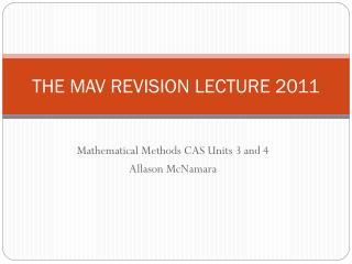
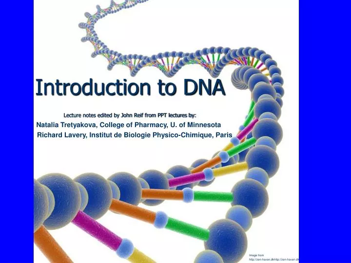
Introduction to DNA Lecture notes edited by John Reif from PPT lectures by:. Natalia Tretyakova, College of Pharmacy, U. of Minnesota. Richard Lavery, Institut de Biologie Physico-Chimique, Paris. Image from http://zen-haven.dkhttp://zen-haven.dk. DNA Double helix

E N D
Introduction to DNA Lecture notes edited by John Reif from PPT lectures by: Natalia Tretyakova, College of Pharmacy, U. of Minnesota Richard Lavery, Institut de Biologie Physico-Chimique, Paris Image from http://zen-haven.dkhttp://zen-haven.dk
DNA • Double helix • Stores genetic code as a linear sequence of bases • ≈ 20 Å in diameter • Human genome ≈ 3.3 x 10⁹ bp • ≈ 25,000 genes Richard Lavery Institut de Biologie Physico-Chimique, Paris
Chemical bond 1 Å (10-10 m) Amino acid 10 Å (10-9 m) Globular protein 100 Å (10-8 m) Virus 1000 Å (10-7 m) Cell nucleus 1 mm (10-6 m) Bacterial cell 5 mm (10-5 m) Chromosome DNA 10 cm (10-1 m) Biological length scale Richard Lavery Institut de Biologie Physico-Chimique, Paris
The Building Blocks of DNA OH ribose H deoxyribose Nucleoside Nucleotide Richard Lavery Institut de Biologie Physico-Chimique, Paris
Nucleotides are linked by phosphodiester bonds • Strand has a direction (5'3') • DNA is negatively charged on phosphate backbone. Richard Lavery Institut de Biologie Physico-Chimique, Paris
C5 C4 C6 N7 C5 C6 N3 C8 N1 N1 C2 N9 C4 C2 N3 Purine (Pur / R) Pyrimidine (Pyr / Y) Base families Richard Lavery Institut de Biologie Physico-Chimique, Paris
DNA and RNA nucleobases • (DNA only) • (RNA only) • Natalia Tretyakova • College of Pharmacy, U. of Minnesota
Purine BasesThe 9 atoms that make up the fused rings (5 carbon, 4 nitrogen) are numbered 1-9. All ring atoms lie in the same plane. Richard B. Hallick Introductory Course in Biology or Biochemistry
Purine Nucleotides • Natalia Tretyakova • College of Pharmacy, U. of Minnesota
Pyrimidine BasesAll pyrimidine ring atoms lie in the same plane. Richard B. Hallick Introductory Course in Biology or Biochemistry
Pyrimidine Nucleotides • Natalia Tretyakova • College of Pharmacy, U. of Minnesota
nucleobase • (Deoxy) • nucleoside • 5’-mononucleotide • Adenine (A) • Guanine (G) • Thymine (T) • Cytosine (C) • Uracil (U) • 2’-Deoxyadenosine (dA) • 2’- Deoxyguanosine (dG) • 2’- Deoxythymidine • (dT) • 2’- Deoxycytidine • (dC) • Uridine (U) • Deoxyadenosine 5’-monophosphate • (5’-dAMP) • Deoxyguanosine 5’-monophosphate • (5’-dGMP) • Deoxythymidine 5’-monophosphate • (5’-dTMP) • Deoxycytidine 5’-monophosphate • (5’-dCMP) • Uridine 5’-monophosphate (5’-UMP) • Nomenclature of nucleobases, nucleosides, and mononucleotides • Natalia Tretyakova • College of Pharmacy, U. of Minnesota
Structural differences between DNA and RNA • DNA • RNA • Natalia Tretyakova • College of Pharmacy, U. of Minnesota
Deoxyribose Sugar The hydroxyl groups on the 5'- and 3'- carbons link to the phosphate groups to form the DNA backbone. Richard B. Hallick Introductory Course in Biology or Biochemistry
Nucleosides • A nucleotide is a nucleoside with one or more phosphate groups covalently attached to the 3'- and/or 5'-hydroxyl group(s). Richard B. Hallick Introductory Course in Biology or Biochemistry
Preferred conformations of nucleobases and sugars in DNA and RNA • Sugar puckers: • 5.9 A • 7.0 A • Natalia Tretyakova • College of Pharmacy, U. of Minnesota
Nucleosides Must Be Converted to5’-Triphosphates to be Part of DNA and RNA • Natalia Tretyakova • College of Pharmacy, U. of Minnesota
Thymine -Adenine Cytosine -Guanine Watson-Crick base pairs Richard Lavery Institut de Biologie Physico-Chimique, Paris
A-T and G-C Base Pairing Richard B. Hallick Introductory Course in Biology or Biochemistry
Hydrogen bond donors and acceptors on each edge of a base pair • Natalia Tretyakova • College of Pharmacy, U. of Minnesota
Purine always binds with a Pyrimidine Richard Lavery Institut de Biologie Physico-Chimique, Paris
Base pair dimensions Richard Lavery Institut de Biologie Physico-Chimique, Paris
DNA : A ,T,G,C + deoxyribose RNA : A,U,G,C + ribose DNA/RNA chemical structure Richard Lavery Institut de Biologie Physico-Chimique, Paris
Helix Axis View: Backbone structure: • Alternating backbone of deoxyribose and phosphodiester groups • Chain has a direction (known as polarity), 5'- to 3'- from top to bottom • Oxygens (red atoms) of phosphates are polar and negatively charged • Bases extend away from chain, and stack atop each other • Bases are hydrophobic Richard B. Hallick Introductory Course in Biology or Biochemistry
Video of DNA Helix Structure: http://www.youtube.com/watch?v=ZGHkHMoyC5I Contains material from: Alberts, Bray, Hopkin, Johnson, Lewis, Raff, Roberts, Walter, Essential Cell Biology, Second Edition, Garland Science Publishing, 2004
B-DNA Structure CGCGTTGACAACTGCAGAATC Richard Lavery Institut de Biologie Physico-Chimique, Paris
Features of the B-DNA Double Helix • Two DNA strands form a helical spiral, winding around a helix axis in a right-handed spiral • The two polynucleotide chains run in opposite directions • The sugar-phosphate backbones of the two DNA strands wind around the helix axis like the railing of a sprial staircase • The bases of the individual nucleotides are on the inside of the helix, stacked on top of each other like the steps of a spiral staircase. Richard B. Hallick Introductory Course in Biology or Biochemistry
B-DNA (axial view) Richard Lavery Institut de Biologie Physico-Chimique, Paris
R.H. helix B-DNA (lateral view) Richard Lavery Institut de Biologie Physico-Chimique, Paris
Base stacking: an axial view of B-DNA • Natalia Tretyakova • College of Pharmacy, U. of Minnesota
Forces stabilizing DNA double helix • Hydrogen bonding (2-3 kcal/mol per base pair) • Stacking (hydrophobic) interactions (4-15 kcal/mol per base pair) • Electrostatic forces. Comparison to other bonds • Covalent Bond Energies: • C-C 85 kcal/mol • C-O 87 kcal/mol • Natalia Tretyakova • College of Pharmacy, U. of Minnesota
B-DNA • •Sugars are in the 2’ endo conformation. • •Bases are the anti conformation. • •Bases have a helical twist of 34.6º (10.4 bases per helix turn) • Helical pitch = 34 A • 23.7 A • right handed helix • helical axis passes through • base pairs • 7.0 A • planes of bases are nearly • perpendicular to the helix axis. • 3.4 A rise between base pairs • Wide and deep • Narrow and deep • Natalia Tretyakova • College of Pharmacy, U. of Minnesota
DNA can deviate from the ideal Watson-Crick structure • Helical twist ranges from 28 to 42° • Propeller twisting 10 to 20° • Base pair roll • Natalia Tretyakova • College of Pharmacy, U. of Minnesota
MAJOR MINOR DNA grooves Richard Lavery Institut de Biologie Physico-Chimique, Paris
Major groove and Minor groove of DNA • N • NH • O • 2 • N • H • N • O • 2 • N • NH • N • N • N • HN • C-1’ • N • N • N • N • C-1’ • NH • O • O • 2 • C-1’ • Hypothetical situation: the two grooves would have similar size if dR residues • were attached at 180° to each other • To deoxyribose-C1’ • C1’ -To deoxyribose • C-1’ • Natalia Tretyakova • College of Pharmacy, U. of Minnesota
N • NH • 2 • H • N • O • 2 • N • N • HN • C-1’ • N • N • NH • O • 2 • C-1’ • Major and minor groove of the double helix • O • N • NH • N • N • N • N • C-1’ • O • C-1’ • Wide and deep • Narrow and deep • Natalia Tretyakova • College of Pharmacy, U. of Minnesota
B-type duplex is not possible for RNA • steric “clash” • Natalia Tretyakova • College of Pharmacy, U. of Minnesota
De-hydration Hydration 3’ 5’ 5’ 3’ Antiparallel strands A B A and B DNA allomorphs Richard Lavery Institut de Biologie Physico-Chimique, Paris
A-DNA (longitudinal view) Richard Lavery Institut de Biologie Physico-Chimique, Paris
R.H. helix A-DNA (lateral view) Richard Lavery Institut de Biologie Physico-Chimique, Paris
A-form helix:dehydrated DNA; RNA-DNA hybrids • •Sugars are in the 3’ endo conformation. • •Bases are the anti conformation. • •11 bases per helix turn • Helical pitch = 25.3 A • Right handed helix • planes of bases are tilted • 20 ° relative the helix axis. • 2.3 A rise between base pairs • 25.5 A • Top View • Natalia Tretyakova • College of Pharmacy, U. of Minnesota
The sugar puckering in A-DNA is 3’-endo • 5.9 A • 7.0 A • Natalia Tretyakova • College of Pharmacy, U. of Minnesota