Download
1 / 66
660 likes | 667 Views
Obstructive uropathy-induced hypervolemia presenting as new-onset Heart failure. Noon Conference Alexis Hill September 3 rd 2019. Outline. 1. Case review 2. Disease pathogenesis 3. Differential diagnosis 4. Diagnostics 5. Treatment 6. Patient update. HPI.

E N D
Obstructive uropathy-induced hypervolemia presenting as new-onset Heart failure Noon Conference Alexis Hill September 3rd 2019
Outline 1. Case review 2. Disease pathogenesis 3. Differential diagnosis 4. Diagnostics 5. Treatment 6. Patient update
HPI • 73 yo Caucasian male presents with chief complaint of increasing SOB over the past two to three months, as well as leg swelling for the last couple of weeks. • “But while I have you here, what do you think of the keto diet? I’ve put on so much weight in the past couple of months and I’m really trying to lose it.
Past Medical History • Admits only to tobacco use and psoriasis • “Well doc, I haven’t been to the doctor’s in 20 years, but I think I’m pretty healthy. The only reason I came in was because I couldn’t get my shoes on this morning.”
Past surgical history • Bilateral hip replacements (2/2 OA)
Family History • Non-contributory
Social history • Tobacco use: cigars (2-4 filtered cigars per day) • Alcohol use: occasional • Illicit drug use: denies • Works for an insurance company • Lives at home with his wife
ROS • Constitutional: Positive for unexpected weight change (weight gain).Negative for activity change. • Eyes: Negative for pain and discharge. • Respiratory: Positive for shortness of breath. Negative for chest tightness. • Cardiovascular: Positive for leg swelling.Negative for chest pain and palpitations. • Gastrointestinal: Negative for abdominal pain, constipation or diarrhea • Genitourinary: Positive for urgency, frequency, and nocturia. Negative for difficulty urinating or hematuria. • Musculoskeletal: Negative for arthralgias and back pain. • Skin: Negative for color change and rash. • Neurological: Negative for dizziness, light-headedness and numbness. • Psychiatric/Behavioral: Negative for agitation and confusion.
Physical Exam • Vitals: BP 156/96 / P 81 / R 16 / SpO2 99% on RA • Constitutional: Morbidly obese • Caucasian male. In no acute distress, non-toxic appearing. Pleasant and cooperative on exam. • Neck: Neck supple. No tracheal deviation present. JVD present. • Cardiovascular: Intact distal pulses. Normal rate. Irregularly irregular rhythm. 2/6 systolic murmur. • Pulmonary/Chest: Effort normal. No respiratory distress. rales in bilateral lung bases. • Abdominal: Soft. Bowel sounds are normal. There is no tenderness. • Musculoskeletal: He exhibits edema(+3 pitting edema bilateral lower extremities up to the knee). • Neurological: He is alert and oriented to person, place, and time. • Psychiatric: He has a normal mood and affect. His behavior is normal.
Imaging: TTE Left ventricle: The cavity size is normal. Wall thickness is mildlyincreased. Systolic function is at the lower limits of normal by thebiplane method of disks. The estimated ejection fraction is 53%. There areno regional wall motion abnormalities. Unable to assess LV diastolicfunction due to atrial fibrillation Aortic valve: There is mild stenosis. There is no significantregurgitation. Mean gradient (S): 15 mm Hg. Peak gradient (S): 23 mm Hg. SVI is a bit low = 29 ml/m2, and the patient is in AF, gradient may be abit underestimated. Dimensionless index: 0.43. Valve area (VTI): 1.6 cm2.
Imaging: RP US The right kidney measures 12.3 x 5.7 x 5.7 cm. There is mild hydronephrosis. No mass or calculus is seen. There is no perinephric fluid. The left kidney measures 12 x 6.6 x 7.1 cm and also demonstrates mild hydronephrosis. A medial cyst measuring 9.1 x 7.8 x 8.3 cm is present. There is no evidence of soft tissue mass or calculus. To the limited extent seen, the bladder appears to be distended and may demonstrate some wall trabeculation.
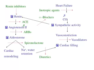
Brief Hospital Course • Patient was admitted to telemetry floor, started on hydralazine and isosorbide dinitrate. Nephrology and cardiology (heart failure) were consulted. • Nephrology – started bumetanide gtt, metolazone. Recommended Foley placement due to concern for component of obstructive uropathy – patient refused. • Cardiology – started heparin gtt (CHA2DS2-VASc Score: 3), added metoprolol tartrate for rate control. Recommended Foley placement due to concern for component of obstructive uropathy – patient refused. Consulted urology for help.
Brief hospital course • Urology - Obtained PVR (350 cc residual post-void). Prostate exam revealed markedly enlarged prostate without nodules or other irregularities. STRONGLY Recommended Foley placement – patient agreed. 2 L UOP immediately after Foley placement. Placed on tamsulosin.
Brief Hospital Course • Put out 14 L (30 pounds of water) overnight after Foley placement – bumetanide gtt and metolazone discontinued. Patient diuresed a total of 23 L of urine over the course of his admission. • His shortness of breath and lower extremity edema completely resolved. • Cr trended down dramatically and actually normalized (7.96 -> 3.57 -> 1.11). • Discharged in stable condition on daily tamsulosin with Foley in place and plans for close outpatient follow-up with urology.
UTO: Epidemiology • A common problem, but actually a rare cause of significant AKI • UTO is a more common cause of renal failure in children due to contribution of congenital abnormalities • Incidence in men > women, particularly with increasing age – primarily due to prostatic hypertrophy • An important cause of renal failure in malignancy
UTO: ETIOLOGY • Obstruction may occur at any point along the genitourinary tract • Causes of obstruction are many – stones, urethral stricture, posterior urethral valves, prostatic hypertrophy, neurogenic bladder (diabetes, spinal cord injuries, MS, etc.), cancer, external compression (fecal impaction, enlarged lymph nodes, retroperitoneal fibrosis), blood clots
UTO: Clinical Presentation • Pain is frequently absent! Especially if slowly progressive obstruction • LUTS common with urethral obstruction (frequency, urgency, nocturia, polyuria) • An enlarged bladder may be detected by percussion of pelvis/lower abdomen, although exam may be difficult in obese patients
UTO: Laboratory findings • Elevated serum creatinine • Typically requires obstruction of bilateral kidneys • Hematuria/pyuria • Hyperkalemic RTA • Possible due to mineralocorticoid resistance with impaired distal Na resorption due to reduced activities of transporter proteins
UTO: Diagnosis • Early diagnosis is critical as most cases can be corrected with appropriate intervention, but delay in action can lead to irreversible renal injury • Diagnosis primarily by imaging – ultrasound preferred modality of choice for practical reasons • Hallmark of obstruction: dilation of collecting system in one or both kidneys • Pitfall: high false positive rate (26%) as mild hydronephrosis may be present without obstruction • Good for ruling outobstruction - negative predictive value 98% • Exception: non-contrast CT should be test of choice if stone is suspected as etiology of obstruction
UTO: Diagnosis • Confirmation: Foley placement • Bladder scans are also useful, but operator dependent and may be limited by body habitus – if in doubt, place the foley
UTO: Treatment • Treatment is straight-forward – treating the cause of the obstruction • In the case of BPH: • Medical therapy (Alpha-1-antagonists usually first line) • Foley placement • Surgical intervention (typically transurethral resection)
Post-obstructive Diuresis • Defined as UOP exceeding 200 cc/hr for > 2 hours or >3 L UOP in 24 hours after obstruction is relieved • Typically a normal physiologic response to help eliminate excess volume and solutes accumulated during obstruction • In most patients, diuresis slows as solute and volume homeostasis is achieved • Considered pathologic when kidneys continue to eliminate salt and water even after homeostasis has been achieved • May lead to dangerous electrolyte imbalances and severe dehydration
Post-obstructive diuresis • No need to “gradually decompress” bladder • However, patients with POD should be admitted for close monitoring of volume and electrolyte status • Fluid replacement: recommended 75% of previous one hour of UOP
Update: How is our patient doing? • Has been seen by urology twice since discharge – unfortunately failed voiding trial at both visits, although his renal function remains completely normal. Currently not interested in TURP – doing well for now with tamsulosin and intermittent straight catheterizations 3x daily. • Most recently seen by our own Dr. Dayal just last week – was doing well at that time, planning upcoming vacation with his wife.
PGY 2 Case Presentation Mary Lareine Pastoral, MD September 3, 2019 29
Outline • Review Case • Discuss Disease Pathogenesis • Discuss Differential Diagnosis • Discuss Diagnostic Criteria • Discuss Treatment • Patient Update 30
History of Present Illness September 2017: First evaluated by Rheumatology. S: 45 year old woman with past medical history of fibromyalgia, with chief complaint of diffuse joint pain of 20 years. O: Hands:R:+2/3 2-3 MCP, 1/3 2nd PIP, 2/3 3rd PIP m tenderness, no erythema, no warmth, no swellingL:+2/3 2-3 PIP, 1,3rd, MCP tenderness, no erythema, no warmth, no swelling Erosive changes on MRI, elevated inflammatory markers Assessment: Seronegative Rheumatoid Arthritis Plan: Started on methotrexate 10 mg once a week, folic acid, prednisone 2.5. December 2017: erosive changes, persistent symptoms- TNF inhibitor (Humira) added May 2018: elevated LFTs ALT 213, AST 90 of note pt was started 2 months prior on Lipitor, methotrexate dose decreased persistent transaminitis methotrexate stopped 6/2018 Summa Health Sample Preso
History of Present Illness February 2019: (+) no vision in right eye when she covered left eye 2 weeks ago (+) occasional right eye pain described as "stretched muscle" discomfort when moving her eyes right and down 1 or 2 times flashes of light, ~5 floaters Physical exam: Summa Health Sample Preso
History of Present Illness February 2019: Admitted to Aultman Hospital for worsening eye symptoms. She was given a five day course of IV steroids. May 2019: Patient started having continuous hemifacial spasms on the left, leg cramps and spasms, lasting for 3 hours. She retrospectively thinks she had previous episodes of hemifacial spasms back in the 2017. She denies a history of Bell's palsy. CSF analysis repeated and showed positive oligoclonal bands. She was given another course of IV steroids for 5 days. She continues to have intermittent facial spasms, occasionally, lasting <1h, as well as intermittent spasms in the legs. Summa Health Sample Preso
History of Present Illness June 2019: Rheumatology visit. Information reviewed. MRIs showed a few non-specific lesions. There were no lesions in the cervical and thoracic cord. CSF analysis was done (without OCB testing initially, showed 43WBC and elevated IgG synthesis rate). She was tested for Aquaporine-4 and MOG IgG and both were negative (sent to Mayo). After receiving the 5 days of IV steroids on May 2019, symptoms improved considerably but incompletely over the next few weeks Humira stopped. Change in treatment to Rituxan planned. July 2019: Rituximab treatment recommended by neurology for management of both multiple sclerosis with optic neuritis as well as Rheumtoid arthritis . Summa Health Sample Preso
History of Present Illness July 2019: evaluated by Neurology, impression was possible TNF-alfa inhibitor-related demyelinating optic neuritis and the recommendation was to initiate Rituximab. August 2019: Started on Rituximab She now has 20/100 VA in this eye and has prominent dyschromatopsia. Summa Health Sample Preso
Past Medical History 37 Fibrolmyalgia Sleep apnea, not using CPAP machine Bipolar disorder, well controlled on Lamictal
Past Surgical History 38 C section THBSO Wrist surgery for Ganglion’s cyst of right wrist
Family History 39 Mother: fibromyalgia, heart disease Father: diabetes, hyperlipidemia, heart disease, arthritis Sister: bipolar disorder
Social history 40 Smoking: former smoker Alcohol: denies alcohol use Illicit drug use: denies
Review of Systems 42 Constitutional: Positive for unexpected weight change(gained 15 lbs since February 2019). Negative for appetite change, chills and fever. HENT: Positive for tinnitus (left). Negative for congestion, hearing loss, mouth sores, nosebleeds, trouble swallowing and voice change. Eyes: Positive for visual disturbance(color blind in right eye, since the optic neuritis) , (+) double vision. Negative for pain and redness. Respiratory: Negative for cough, chest tightness, shortness of breath and wheezing. Cardiovascular: Negative for chest pain and palpitations. Gastrointestinal: Negative for abdominal pain, blood in stool, constipation, diarrhea, nausea and vomiting. Endocrine: Positive for heat intolerance. Genitourinary: Negative for difficulty urinating, dysuria and hematuria. Musculoskeletal: Positive for arthralgias, back pain, myalgias, neck pain and neck stiffness. Negative for joint swelling. Skin: Negative for rash. Neurological: Positive for dizziness, tremors, light-headedness (+) some numbness over at the left index finger. Negative for headaches. Psychiatric/Behavioral: Positive for dysphoric mood.
Physical exam 44 BP 112/78 | Pulse 97 | Resp 18 | Ht 172.7 cm (5' 8") | Wt 83.3 kg (183 lb 9.6 oz) | BMI 27.92 kg/m² HEENT: Visual acuity to near card was as follows: OD= 20/100 (with glasses) OS= 20/25 (with glasses).Visual fields were full to confrontation.Pupils were 4 mm, less reactive with a relative afferent pupillary defect in OD.Funduscopic examination was normal without disc edema, erythema, or atrophy. Ocular ductions were full without nystagmus or ataxia. Neck: No thyromegaly CV: Normal rate and regular rhythm. Thorax: Clear breath sounds Abdomen: soft, non-tender, no organomegaly Neuro: Sensation intact, reflexes normal, Coordination testing in the arms and legs was performed including point-to-point, rapid-alternating, and fine movements. Rapid movements were smooth with good cadence and there was no dysmetria or ataxia. No signs of cerebellar dysfunction. Gait was normal, tandem walking was impaired. Extremities: see next slide
Musculoskeletal:Neck: no tenderness to palpationSpine: no tenderness to palpationShoulders: intact ROM, no effusion, no tenderness, no erythema, no warmth, no swellingElbows: intact ROM, no effusion, no tenderness, no erythema, no warmth, no swellingWrists: R:no effusion, (+) tenderness, no erythema, no warmth, no swellingL:no effusion, (-)tenderness, no erythema, no warmth, no swellingHands:R: (+) 2nd-4th MCP, 2nd PIP tenderness, no erythema, no warmth, no swellingL:no tenderness, no erythema, no warmth, no swellingHips: negative log rollKnees: no effusion, no tenderness, no erythema, no warmth, no swellingAnkles: no effusion, no tenderness, no erythema, no warmth, no swellingFeet:R:no tenderness, no erythema, no warmth, no swellingL:no tenderness, no erythema, no warmth, no swellingGait: No antalgic gait Summa Health Sample Preso
Laboratory tests Summa Health Sample Preso
Laboratory tests Aquaporin-4 and myelin oligodendrocyteglycoprotein MOG-IgG: reported negative (sent to Mayo)CSF (February 2019): WBC 43, RBC 0, protein and glucose normal, IgG synthesis rate 19.2 (normal 0-12) CSF (May 2019): positive oligoclonal band, increased WBC and a traumatic tap. Summa Health Sample Preso
Imaging Brain MRI June 19, 2019: non specific white matter abnormalities, no enhancing lesions stable compared to February 2019 Cervical and thoracic cord MRI June 19, 2019: normal cord, no leptomeningeal enhancement 49