Download
1 / 36
360 likes | 705 Views
Case Presentation. 31MNo PMHxSingle GSW to neckHandgun at 15 ft range. Case Presentation. Hemodynamically stable130/75, 86, 18, 99% on NC Spontaneous, regular respirations. Exam. 0.5 cm entry wound at midline in Zone INo exit woundSmall hematoma, no active bleedingNo crepitusBreath sounds

E N D
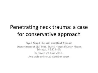
1. Penetrating Neck Trauma Umut Sarpel
PGY-4
2. Case Presentation 31M
No PMHx
Single GSW to neck
Handgun at 15 ft range
3. Case Presentation Hemodynamically stable
130/75, 86, 18, 99% on NC
Spontaneous, regular respirations
4. Exam 0.5 cm entry wound at midline in Zone I
No exit wound
Small hematoma, no active bleeding
No crepitus
Breath sounds b/l
2+ pulses b/l UE
4/4 strength, sensation b/l
CN II-XII grossly intact
5. Management Labs - Hct 44
otherwise unremarkable
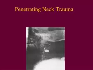
CXR obtained
7. Management CXR: R hemothorax
R chest tube ? 500 cc blood
Flexible laryngoscopy ? no obvious injury
Airway control ? fiberoptic awake nasotracheal intubation by anesthesia
8. Management
Angiogram obtained:
13. Operative Course Median sternotomy
Pseudoaneurysm of brachiocephalic artery
Proximal/distal control
Interposition graft with PTFE from brachiocephalic to subclavian artery
14. Operative Course Injury to brachiocephalic vein noted; controlled and ligated
Neck dissection ? no tracheal injury
Rigid esophagoscopy ? no injury noted
15. Post-Op Course Post-op head CT: no infarct
SICU: ventilatory support
Moderate output from chest tube
2U PRBC on POD#3
Neurologically intact
Progressive vent weaning
16. Overview Complex anatomy, many organ systems,
each requiring evaluation:
Vascular
Respiratory
Digestive
Neurologic
Endocrine
Skeletal
18. Overview Anatomy
Signs / symptoms of injury
Evaluation
Management
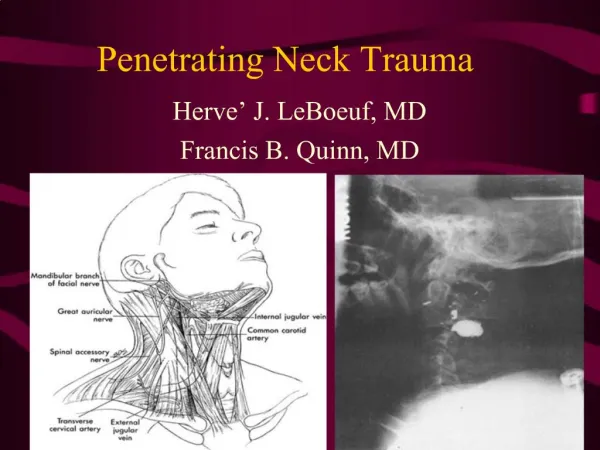
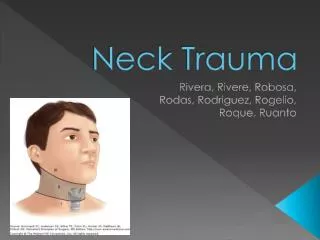
19. Anatomy: Zones
20. Anatomy: Zones Zone I � inferior trachea and esophagus vessels of the root of the neck: the brachiocephalic trunk, the subclavian arteries, the common carotid arteries, the thyrocervical trunk and the corresponding veins, thoracic duct, thyroid gland, spinal cord.
Zone II � the larynx, hypopharynx common carotid arteries the internal and external carotid arteries the internal jugular veinsand cranial nerves 10, 11, and 12, the spinal cord.
Zone III � the pharynx carotid arteries, the vertebral arteries, the internal jugular veinsZone I � inferior trachea and esophagus vessels of the root of the neck: the brachiocephalic trunk, the subclavian arteries, the common carotid arteries, the thyrocervical trunk and the corresponding veins, thoracic duct, thyroid gland, spinal cord.
Zone II � the larynx, hypopharynx common carotid arteries the internal and external carotid arteries the internal jugular veinsand cranial nerves 10, 11, and 12, the spinal cord.
Zone III � the pharynx carotid arteries, the vertebral arteries, the internal jugular veins
21. Signs: Vascular Injury Shock
Hemorrhage
Hematoma
Evolving stroke
Pulse differential in upper extremities
Bruit or thrill
22. Signs: Laryngotracheal Injury Subcutaneous emphysema
Sucking wound
Hemoptysis
Dyspnea
Stridor
Hoarseness or dysphonia
23. Signs: Esophageal Injury Often clinically silent
Milder subcutaneous emphysema
Bloody saliva
Dysphagia or odynophagia
Fever (late)
24. Signs: Spinal Injury Neurologic defect
Spinal shock
Hypotensive, often not tachycardic
(But in a hypotensive trauma pt,
always assume hemorrhagic shock first)
25. Mechanism Stab wound
What you see is what you get
GSW
Unpredictable trajectory
Thermal injury
Maintain high level of suspicion
26. Table 1 �
Mortality has decreased over the years
Most still due to exsanguination
Table 2 � McConnel paper combined data from 1963-1990 papers (2,495 pts)
Most common sight of injury is aerodigestive tract (20%)
IJ was the most commonly injured vessel followed by carotid.Table 1 �
Mortality has decreased over the years
Most still due to exsanguination
Table 2 � McConnel paper combined data from 1963-1990 papers (2,495 pts)
Most common sight of injury is aerodigestive tract (20%)
IJ was the most commonly injured vessel followed by carotid.
27. Evaluation Old standard: formal neck exploration for all penetrating trauma that violates platysma
Was a/w 50% negative exploratory rate
New focus on directed exams: angiography, esophagoscopy, esophagography, laryngoscopy
28. Management: Vascular Injuries Zone II vascular injuries readily apparent
Zone I and III injuries more difficult to detect due to anatomical constraints:
32% of pts w/ major Zone I vascular injury had
no localizing PE findings
29. Management: Vascular Injuries Angiography: adjunctive diagnostic tool
Arteriogram can also be therapeutic w/ embolization (works esp well in Zone III where vessels are smaller)
Duplex exam:
in qualified centers may be acceptable
alternative
30. Management: Vascular Injuries In general, vessels should be repaired rather than ligated
Carotid injuries should be repaired unless there is an already established dense neurologic deficit w/ edema (revascularization may convert ischemic to hemorrhagic infarct)
If bypass is needed, PTFE preferred over saphenous vein graft
32. Management: Esophageal Injury Early detection of injury is paramount
If repaired < 24hrs, survival 90%
If > 24 hours, survival 64%
Best detected by combination of esophagoscopy and esophagography (sensitivity near 100%)
Rigid / flexible endoscopy both acceptable
33. Management: Esophageal Injury Operative repair:
Primary closure is ideal (esp < 24 hrs)
Close over a T-tube
Buttress w/ muscle flaps or pleura
Divert with esophageal stoma
Widely drain
Fistula rate up to 57%
Consider routine swallow studies
34. Management: Tracheal Injury Thorough laryngoscopy
Primary repair is the rule, tracheal mobility allows closure of defects up to 2-3cm
Tracheotomy rarely indicated, only for a large defect (increases risk of infection)
Absorbable suture Entry incision in the cricoid cartilage, extend in the midline to the thyroid membrane, and meticulously close mucosal lacerations, using advancement flaps if necessary, or rarely, grafts. Wire vs. miniplate fixation of cartilaginous fractures.Entry incision in the cricoid cartilage, extend in the midline to the thyroid membrane, and meticulously close mucosal lacerations, using advancement flaps if necessary, or rarely, grafts. Wire vs. miniplate fixation of cartilaginous fractures.
35. Management: Spinal Injury Can only prevent further injury
Steroids
appear to have some benefit in blunt
trauma, but no evidence for routine use
in penetrating trauma
36. Algorithm A large amount of literature accumulated showing mandatory exploration is not always necessary.A large amount of literature accumulated showing mandatory exploration is not always necessary.
37. Conclusions Know your anatomy
Neck exploration is no longer mandatory in asymptomatic pts
Physical exam is probably the most useful diagnostic tool (esp Zone II)
Non-invasive diagnostic / therapeutic modalities should be utilized