Download
1 / 57
600 likes | 902 Views
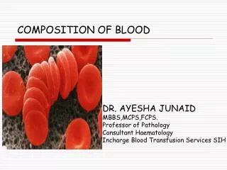
BLOOD COMPOSITION. dr. Husnil Kadri , M.Kes Biochemistry Departement Medical Faculty Of Andalas University Padang. BIOSINTESIS HEMOGLOBIN (PORFIRIN). Struktur Porfirin. Porfirin adalah senyawa siklik yg dibentuk oleh 4 cincin pirol.

E N D
BLOOD COMPOSITION dr. HusnilKadri, M.Kes Biochemistry Departement Medical Faculty Of Andalas University Padang
Struktur Porfirin • Porfirin adalah senyawa siklik yg dibentuk oleh 4 cincin pirol. • Masing-masing cincin dihubungkan oleh 4 jembatan metenil (-HC=). • Sifat khas porfirin adalah atom nitrogennya mampu mengikat ion logam. • Contoh; - heme pada Hb mengikat Fe - klorofil pada tumbuhan hijau mengikat Mg
Beberapa Hemoprotein Protein Fungsi - Hemoglobin mengangkut oksigen di dalam darah - Mioglobin menyimpan oksigen di dalam otot - Sitokrom c keterlibatan pada rantai transpor elektron - Sitokrom P450 hidroksilasi xenobiotik - Katalase degradasi hidrogen peroksida - Triptofan pirolase oksidasi triptofan
SintesisHemediMitokondria • 85% sintesis heme terjadi dalam sel pembentuk eritrosit pada sumsum tulang • Heme disintesis dari suksinil KoA + glisin. • Piridoksal fosfat diperlukan untuk mengaktifkan glisin.
Pengaturan Sintesis Heme • Enzim regulator adalah ALA-sintase. • Heme bertindak sebagai regulator negatif (umpan balik negatif) sintesis enzim ALA- sintase. • Jika heme meningkat, maka sintesis ALA-sintase akan menurun.
Porfiria • Merupakan gangguan genetik biosintesis heme. • Umumnya autosomal dominan, kecuali porfiria eritropoitik kongenital. • Gejala; - nyeri abdomen - gangguan neuropsikiatri - fotosensitifitas kulit - bila berat = prototipe manusia srigala
Dasar Biokimia Porfiria Mutasi DNA Abnormalitas enzim pada sintesis heme Akumulasi ALA & PBG atau Akumulasi porfirinogen penurunan heme dlm sel & di kulit & jaringan tubuh Cairan tubuh Tanda &gejala Oksidasi spontan porfirinogen neuropsikiatrik menjadi porfirin Fotosensitifitas
Terapi Porfiria • Hanya simptomatik. • Represor ALA-sintase; - glukosa - hematin (bentuk hidroksida dari heme) - b-karoten untuk fotosensitifitas - preparat tabir surya • Kontraindikasi; - preparat anestesi - alkohol - griseofulvin & barbiturat
Fungsi Utama Darah 1. Respirasi; pengangkutan O2 dan CO2 2. Nutrisi; pengangkutan hasil absorpsi usus 3. Ekskresi; pengangkutan sisa metabolik ke ginjal, paru-paru, kulit, & usus
Fungsi Utama Darah 4. Keseimbangan asam-basa 5. Keseimbangan air; antara sirkulasi darah dan jaringan 6. Pengaturan suhu tubuh 7. Pertahanan terhadap infeksi; oleh sel darah putih & antibodi
Fungsi Utama Darah 8. Pengangkutan hormon & pengaturan metabolisme 9. Pengangkutan metabolit 10. Koagulasi
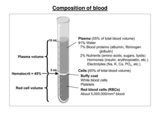
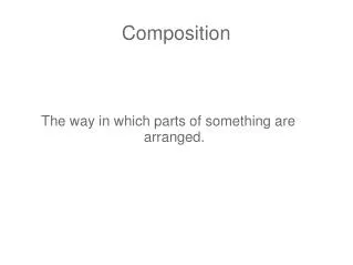
Components of Whole Blood Plasma(55% of whole blood) Buffy coat:leukocyctes and platelets(<1% of whole blood) Formed elements Erythrocytes(45% of whole blood) Withdraw blood and place in tube Centrifuge 1 2 • Hematocrit • Males: 47% ± 5% • Females: 42% ± 5%
Physical Characteristics of Blood • Average volume of blood: • 5–6 L for males; 4–5 L for females (Normovolemia) • Hypovolemia - low blood volume • Hypervolemia - high blood volume • Viscosity (thickness) - 4 - 5 (where water = 1) • The pH of blood is 7.35–7.45; x = 7.4 • Osmolarity = 300 mOsm or 0.3 Osm • This value reflects the concentration of solutes in the plasma • Salinity = 0.85% • Reflects the concentration of NaCl in the blood • Temperature is 38C, slightly higher than “normal” body temperature • Blood accounts for approximately 8% of body weight
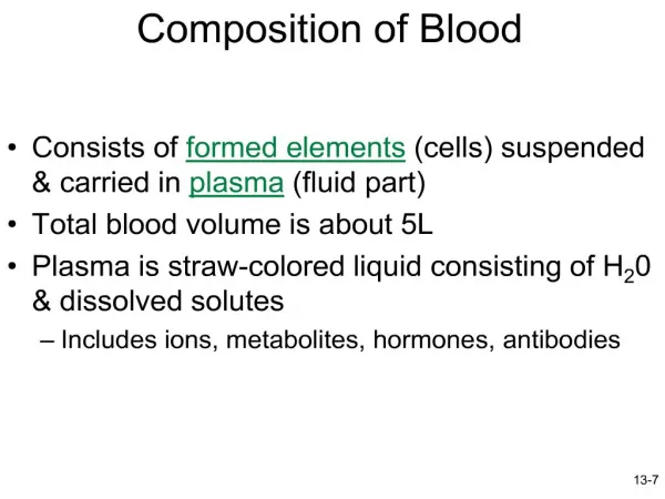
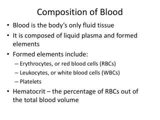
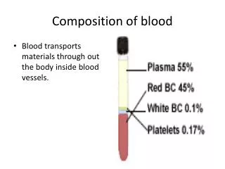
Components of Blood • 55% plasma • 45% cells • 99% RBCs • < 1% WBCs and platelets
Blood Plasma • Blood plasma components: • Water = 90-92% • Proteins = 6-8% • Organic nutrients – glucose, carbohydrates, amino acids • Electrolytes – sodium, potassium, calcium, chloride, bicarbonate • Nonprotein nitrogenous substances – lactic acid, urea, creatinine • Respiratory gases – oxygen and carbon dioxide
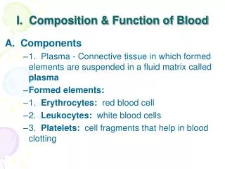
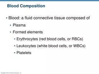
Formed Elements • Formed elements comprise 45% of blood • Erythrocytes, leukocytes, and platelets make up the formed elements • Only WBCs are complete cells • RBCs have no nuclei or organelles, and platelets are just cell fragments • Most blood cells do not divide but are renewed by cells in bone marrow
Erythrocytes (RBCs) • Biconcave disc • Folding increases surface area (30% more surface area) • Plasma membrane contains spectrin • Give erythrocytes their flexibility • Anucleate, no centrioles, no organelles • End result - no cell division • No mitochondria means they generate ATP anaerobically • Prevents consumption of O2 being transported • Filled with hemoglobin (Hb) - 97% of cell contents • Hb functions in gas transport • Hb + O2 HbO2 (oxyhemoglobin) • Most numerous of the formed elements • Females: 4.3–5.2 million cells/cubic millimeter • Males: 5.2–5.8 million cells/cubic millimeter
Erythrocytes (RBCs) Figure 17.3
Erythrocyte Function • Erythrocytes are dedicated to respiratory gas transport • Hemoglobin reversibly binds with oxygen and most oxygen in the blood is bound to hemoglobin • Composition of hemoglobin • A protein called globin • made up of two alpha and two beta chains • A heme molecule • Each heme group bears an atom of iron, which can bind to one oxygenmolecule • Each hemoglobin molecule thus can transport four molecules of oxygen
Structure of Hemoglobin Figure 17.4
Hemoglobin • Satu mol. Hb dewasa (HbA) mempunyai; - 4 gugus heme - Setiap heme mengandung 1 ion Fe2+ - 4 subunit protein globin - Setiap subunit mengikat 1 mol. O2 - 1 mol. Globin mengikat 1 mol. CO2 • Subunit rantai terdiri dari 2 a dan 2 b; - a masing-masing=141 asam amino - b masing-masing = 146 asam amino
Hemoglobin • Oxyhemoglobin – hemoglobin bound to oxygen • Oxygen loading takes place in the lungs • Deoxyhemoglobin – hemoglobin after oxygen diffuses into tissues (reduced Hb) • Carbaminohemoglobin – hemoglobin bound to carbon dioxide • Carbon dioxide loading takes place in the tissues
WBC Anatomy and Types • All WBCs (leukocytes) have a nucleus and no hemoglobin • Granular or agranular classification based on presence of cytoplasmic granules made visible by staining • granulocytes are neutrophils, eosinophils or basophils • agranulocytes are monocyes or lymphocytes
Differential WBC Count • Detection of changes in numbers of circulating WBCs (percentages of each type) • indicates infection, poisoning, leukemia, chemotherapy, parasites or allergy reaction • Normal WBC counts • neutrophils 60-70% (up if bacterial infection) • lymphocyte 20-25% (up if viral infection) • monocytes 3 - 8 % (up if fungal/viral infection) • eosinophil 2 - 4 % (up if parasite or allergy reaction) • basophil <1% (up if allergy reaction or hypothyroid)
Neutrophils (Granulocyte) • Polymorphonuclear Leukocytes or Polys • Nuclei = 2 to 5 lobes connected by thin strands • older cells have more lobes • young cells called band cells because of horseshoe shaped nucleus (band) • Fine, pale lilac practically invisible granules • Diameter is 10-12 microns • 60 to 70% of circulating WBCs
Eosinophils (Granulocyte) • Nucleus with 2 or 3 lobes connected by a thin strand • Large, uniform-sized granules stain orange-red with acidic dyes • do not obscure the nucleus • Diameter is 10 to 12 microns • 2 to 4% of circulating WBCs
Basophils (Granulocyte) • Large, dark purple, variable-sized granules stain with basic dyes • obscure the nucleus • Irregular, s-shaped, bilobed nuclei • Diameter is 8 to 10 microns • Less than 1% of circulating WBCs
Lymphocyte (Agranulocyte) • Dark, oval to round nucleus • Cytoplasm sky blue in color • amount varies from rim of blue to normal amount • Small cells 6 - 9 microns in diameter • Large cells 10 - 14 microns in diameter • increase in number during viral infections • 20 to 25% of circulating WBCs
Lymphocytes • B cells - responsible for humoral immunity • T cells - responsible for cell mediated immunity • B cells responsible for production of antibodies • Receptor matches antigen • Cells multiply • Antibodies
T cells • Cytotoxic T cells (Killer T cells) • Bind to cytotoxic cells (eg infected by virus) • Swell • Release toxins into cytoplasm • Helper T cells • Most numerous • Activate B cells, killer T cells • Stimulate macrophages • Suppressor T cells • Regulate activities of other cell types
Monocyte (Agranulocyte) • Nucleus is kidney or horse-shoe shaped • Largest WBC in circulating blood • does not remain in blood long before migrating to the tissues • differentiate into macrophages • fixed group found in specific tissues • alveolar macrophages in lungs • kupffer cells in liver • wandering group gathers at sites of infection • Diameter is 12 - 20 microns • Cytoplasm is a foamy blue-gray • 3 to 8% o circulating WBCs
UNSUR SELULAR DALAM RESPON IMUN • Jalur limfoid yang membentuk limfosit dan subsetnya • Jalur mieloid yang membentuk sel-sel fagosit mononuklear & polimorfonuklear (PMN). PMN terdiri dari: neutrofil, eosinofil, basofil
Platelets • Platelets are fragments of mega-karyocytes • Their granules contain serotonin, Ca2+, enzymes, ADP, and platelet-derived growth factor (PDGF) • Platelets function in the clotting mechanism by forming a temporary plug that helps seal breaks in blood vessels • Platelets not involved in clotting are kept inactive by Nitric Oxide (NO) and prostaglandins
Protein Plasma - Bagian utama unsur padat dalam plasma. - Konsentrasi total protein plasma + 7-7,5 g/dl. - Berbagai protein plasma dapat dipisahkan menurut karakteristik kelarutannya. - Metode pemisahan tsb antara lain; 1. Salting-out (Na2SO4 23%, dll) 2. Elektroforesis
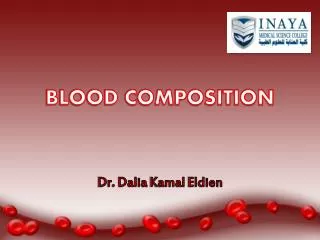
Zone Electrophoresis of Plasma Proteins + - globulins albumin g b a1 a2 pI 6.0 5.6 5.1 4.7
Protein Plasma • Sebagian besar disintesis di hepar. • Umumnya disintesis sbg preprotein pada poliribosom terikat membran. Preprotein akan mengalami modifikasi pascatranslasi. • Hampir semuanya berupa glikoprotein, kecuali albumin. • Bersifat polimorfisme (ciri bawaan pd populasi dgn sedikitnya 2 macam fenotipe). contoh; gol. Darah ABO
Plasma Proteins • More than 200 • Most abundant • Albumin - 4-5 g/100 mL - g-globulins - ~1 g/100 mL • fibrinogen - 0.2-0.4g/100 mL
Albumin - Merupakan protein utama dalam plasma. - Mempertahankan 75-80% tekanan osmotik. - Berfungsi mengikat berbagai macam ligand, seperti; asam lemak bebas, Ca, Cu, Zn, hormon steroid, bilirubin, metheme
Albumin - Albumin juga dapat mengikat obat-an, seperti; sulfonamid, penisilin-G, dikumarol, aspirin - Penyakit hepar akan memperlihatkan rasio albumin/globulin yang menurun.
Transferin • Adalah b1-globulin berbentuk glikoprotein yang disintesis di hepar. • Berfungsi sebagai alat transpor besi (Fe3+) untuk dibawa ke jaringan. • Jika besi tidak diikat oleh transferin, maka akan menjadi prooksidan.
Ceruloplasmin • Protein ini adalah a2-globulin yang mengandung 90% Cu plasma. Tetapi 10% Cu terikat longgar pd albumin, sehingga mudah dilepas ke jaringan. • Ceruloplasmin mengandung ferroksidase yang mengkatalisis ion Fe2+ --> Fe3+, karena hanya ion Fe3+ yang mampu berikatan dgn apotransferin.
g-Globulins • 20% of plasma proteins • “g” refers to electrophoretic mobility • Represents a group of proteins of variable structure • immunoglobulins • Main functional task is immunochemical • Antibodies - combine with specific antigens
Imunoglobulin Plasma • Disintesis dalam sel plasma. • Sel plasma adalah turunan Sel-b yang mensintesis dan mensekresikan imuno- globulin sebagai respon terhadap pajanan berbagai antigen. • Semua imunoglobulin mengandung paling kurang 2 rantai ringan dan 2 rantai berat.
Classes of Immunoglobulins • IgG – Identifies microorganisms for engulfment or lysis • IgE – Inhibits parasite invasion; involved in allergic reactions • IgD – Unknown • IgA – Basis for passive immunity provided by breast milk, agglutinates infectious agents in secretions outside the body, present in tears, mucous • IgM – Identifies microorganisms for engulfment or lysis