Download

1 / 38
380 likes | 582 Views
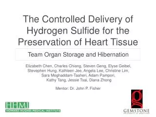
The Controlled Delivery of Hydrogen Sulfide for the Preservation of Heart Tissue. Team Organ Storage and Hibernation. Elizabeth Chen, Charles Chiang, Steven Geng , Elyse Geibel , Stevephen Hung, Kathleen Jee , Angela Lee, Christine Lim, Sara Moghaddam-Taaheri , Adam Pampori ,

E N D
The Controlled Delivery of Hydrogen Sulfide for the Preservation of Heart Tissue Team Organ Storage and Hibernation Elizabeth Chen, Charles Chiang, Steven Geng, Elyse Geibel, Stevephen Hung, Kathleen Jee, Angela Lee, Christine Lim, Sara Moghaddam-Taaheri, Adam Pampori, Kathy Tang, Jessie Tsai, Diana Zhong Mentor: Dr. John P. Fisher
Overview • Introduction • Organ Shortage • Current Methods of Preservation • Background • Ischemia Reperfusion Injury • Hydrogen sulfide attenuates injury • Research Question • Methodology • Results • Conclusions
Organ Shortage • 100,000 patients on organ transplant waiting list • Only 77 patients receive transplants daily • Heart preservation limited to 4-6 hours http://singularityhub.com/2009/06/17/a-look-at-heart-transplants/
Our Goal • Develop a strategy for increasing the viability of stored organs and thus improving patient outcomes
Current Organ Storage Methods • Continuous perfusion • Organ Care System • Effective but expensive • Static cold storage • University of Wisconsin solution • Lack of blood flow leads to I/R injury http://www.news.wisc.edu/newsphotos/uwsolution.html
Na+ Na pump ROS O2 Calcium pump Ca2+ Cold Ischemia Leads to I/R Injury Cardiomyocyte • Continued metabolism • ATP depletion • Accumulation of metabolic waste products • Acidosis Continued cell processes ATP • Ionic balance disruption • Less active ionic pumps • Na+ and Ca2+ accumulate • Cell swelling Lactate, hypoxanthine Mitochondria • ROS production • Inefficiencies in electron transport chain lead to ROS Adapted from: Di Lisa et. al 2007, Jamieson et. al 2008
O2 ROS protons Reperfusion Exacerbates Injury Cardiomyocyte • ROS Burst • Waste products fuel ROS generation Release of cyto c ATP Mitochondria • Mitochondrial Permeability Transition Pore Opens • Protons leak out, no ATP generation Adapted from: Di Lisa et. al 2007, Jamieson et. al 2008
Our Solution: Hydrogen Sulfide (H2S) • H2S • Colorless, poisonous gas • Endogenously produced by cells • Plays critical role in vasoregulation • NaHS is a precursor of H2S • Recent studies • Induced suspended animation in mice1 • Improved left ventricular developed pressure (LVDP)2 • Preserved ATP levels, reduced infarct size3 Molecular structure of H2S 1. Blackstone et al. 2005 2. Li et al. 2007 3. Sivarajahet al. 2006
K+ H2S H2S ROS O2 H2S Protects Hearts from I/R Injury During Ischemia Cardiomyocyte • ROS-scavenging • Directly neutralizes oxygen free-radicals • Upregulates anti-oxidant defenses Ca2+ Dy Mitochondria • mitoK-ATP channel opening • Dissipates ion gradient, • lower Ca 2+ influx H2S Mitochondria • Suspended animation • Reduce metabolic rate • Preserve energy stores • Reduce byproducts Adapted from: Elrod et. al 2007, Hu et. al 2007, Johansen et. al 2006
H2S Depletion [H2S] (μM) Time (min) • H2S depletes from solution over time
Microspheres: A Method for H2S Delivery • Gelatin polymer networks • Means of controlled drug delivery • Can control crosslinkage and loading concentration • Sustain levels of H2S release • Microspheres <10 µm do not cause clots1 http://blogs.indium.com/blog/jim-hisert/microspheres-for-mems 1. Hoshino et al. 2006
Research Question • How can H2S be safely and effectively delivered to prolong organ storage? • Hypothesis • A controlled drug delivery method can sustain H2S levels in the heart and induce protective effects
Objectives • Develop gelatin microspheres for controlled release of H2S • Determine the effects of H2S on rat cardiomyocytes • Determine the efficacy of sustained H2S on rat hearts
Objective 1 • Develop gelatin microspheres for controlled release of H2S • Effect of varying crosslinkage • Effect of varying loading concentration
Microsphere Fabrication Method 1) Fabricate microspheres (vary crosslinkage) Microspheres 2) Load microspheres with NaHS 4) Read absorbance 3) Zinc acetate assay
Microsphere Size Distribution Microspheres less than 10 μm can be fabricated n=144
Effects of NaHS Loading Concentration Uptake of H2S by microspheres increases with loading concentration
Release of H2S by Microspheres Relative H2S levels Time (min) Microspheres enable controlled release of H2S
Objective 2 • Determine the effects of H2S on rat cardiomyocytes • Effect of H2S on cell viability • Effect of H2S on cell metabolism
MTT Assay Method 1) Add NaHS to H9c2 cells 2) Add MTT reagent to media 3) Add MTT solubilizing solution 4) Read absorbance
Effects of H2S on Metabolic Activity Relative absorbances [H2S] (μM) Incubation with 10,000 μM H2S increases metabolic activity
Method: Live-Dead Assay 1) Add microspheres to H9c2 cells 3) Count live cells 2) Add stains Live Dead http://www.invitrogen.com/site/us/en/home/Products-and-Services/Applications/Cell-Culture/primary_cell_culture/Neuronal-Cell-Culture/rat-cortex-and-hippocampus-neurons.html 1) Add microspheres + NaHS to H9c2 cells
Effects of NaHS on Cell Viability Mass of microspheres (mg) Addition of 250mg NaHS-loaded microspheres may improve cell viability
Objective 3 • Determine the efficacy of sustained H2S on rat hearts • Hematoxylin and eosin (H&E) • Caspase-3 • ATP
Surgical Method • Sprague-Dawley rats anesthetized with ketamine and xylazine • Abdominal midline incision • Heparin injected into inferior vena cava prior to exsanguination • Cardioplegia induced • Heart was cooled with saline • UW solution injected into proximal ascending aorta • Vessels were ligated and cut
Tissue Treatment Method Control groups • C-frozen: frozen immediately after explantation • C-ischemia+UW: warm ischemia prior to storage • C-UW: University of Wisconsin (UW) solution
Tissue Treatment Method Experimental groups • E-UW+NaHS: UW solution with 25 mM NaHS • E-UW+S: saline-loaded microspheres • E-UW+S+NaHS: microspheres soaked in NaHS solution NaHS in UW solution NaHS in UW solution NaHS in UW solution NaHS-loaded microspheres PBS-loaded microspheres E-UW+NaHS E-UW+S+NaHS E-UW+S
Histology - H&E • Frozen tissue samples were sliced to 6 µm-thick sections on a cryostat • H&E Stain • Visualize morphology of tissue sample • Hematoxylin: stains nucleic acids blue-purple • Eosin: stains proteins pink • Reveal tissue damage, inflammation
H&E Staining of Rat Heart Tissue 100 μm C-frozen C-UW C-ischemia+UW E-UW+NaHS E-UW+SE-UW+S+NaHS Neither H2S nor microspheres produce a significant inflammatory response
Histology - Caspase-3 • Caspase-3 • A key protein activated in the early stages of apoptosis, or cell death • Utilize an immunoenzymatic reaction to visualize caspase-3
100 μm C-frozen C-UW C-ischemia+UW Caspase-3 Stain of Rat Heart Tissue E-UW+NaHS E-UW+SE-UW+S+NaHS Neither H2S nor microspheres increase apoptosis expression
ATP Assay Method • Frozen samples of left ventricular tissue • ATP Colorimetric/Fluorometric Assay Kit (Abcam, Cambridge, MA) • ATP content assessed at 3 timepoints • ATP content calculated as mM/mg
ATP Concentration as a Measure of Tissue Viability • ATP concentration reflects the hearts energy reserve • The heart especially depends on ATP content, as opposed to other organs • Maintenance of contractile function following storage 1 • ATP content correlated with heart function after reperfusion 2,3 • Hegge, Southard, & Haworth, 2001 • Wang et al., 1991 • Peltz, 2005
Effect of Storage Method on ATP Expression ATP concentration decreases over time
Effect of Storage Method on ATP Expression H2S prolongs ATP preservation
Conclusions • Fabricated microspheres in desired size range • Microspheres yield sustained release of H2S • Released levels of H2S are not harmful to heart cells • H2S prolongs ATP preservation • No significant differences in tissue damage with H2S or microsphere treatment
Future Directions • Alternate measures of in vivo effects • Quantitative apoptosis measures • Functional recovery with reperfusion • Test the system on a larger mammalian subject • Ex. Swine • Evaluate effects on different organs
Acknowledgements • The Gemstone Program • Howard Hughes Medical Institute (HHMI) • Dr. Fisher’s Lab • Mr. Bob Kackley • Mr. Tom Harrod • Dr. Agnes Azimzadeh • Dr. Svetla Baykoucheva • Mr. Chao-Wei Chen • Dr. Nancy Lin • Dr. Ian White • Mr. Andrew Yeatts