Download
1 / 35
400 likes | 517 Views
BASIC Laparoscopy. BY DR/ Mena Zarif Lecturer of general and laparoscopic surgery Faculty of Medicine Sohag university. History of Laparoscopy. A three bladed speculum was found in the ruins of Pompeii*. *A roman town buried by a volcano eruption near modern Naples, Italy - 79 AD).

E N D
BASIC Laparoscopy BY DR/ Mena Zarif Lecturer of general and laparoscopic surgery Faculty of Medicine Sohag university
History of Laparoscopy A three bladed speculum was found in the ruins of Pompeii*. *A roman town buried by a volcano eruption near modern Naples, Italy - 79 AD). The first description dates to Hippocrates in Greece, for use of a speculum to visualize the rectum (460–375 BC).
Hans Christian Jacobaeus(1879 – 1937) • 1910: Swedish internist; first thoracoscopic diagnosis with a cystoscope in a human subject. • Treatment of a patient with tubercular intra-thoracic adhesions. The Possibilities for Performing Cystoscopy in Examinations of Serous Cavities. Münchner Medizinischen Wochenschrift, 1911
Bertram Bernheim • 1911 : First laparoscopy at Johns Hopkins • 12mm proctoscope into epigastric incision on one of Halstead’s patients to stage pancreatic cancer • Bernheim called his procedure ‘organoscopy’ • Findings confirmed on laparotomy
History of Laparoscopy • 1985: Dr. Muhe (Prof Dr Med - Böblingen, Germany)performed the first successful laparoscopic cholecystectomy in a human. However, this was not well publicized until years later. The German Surgical Society rejected Mühe in 1986 after he reported that he had performed the first laparoscopic cholecystectomy.
Minimal access surgery • Minimal access surgery is a marriage of modern technology and surgical innovation that aims to accomplish surgical therapeutic goals with minimal somatic and psychological trauma
Extent of minimal access surger • Laparoscopy • Thoracoscopy • Endoluminal endoscopy • Arthroscopy
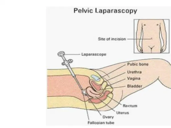
DIAGNOSIS Gallstone Appendicitis Hernia Adhesions Perforated ulcer Hiatus Hernia OPERATION Cholecystectomy Appendicectomy Hernia repair Division of adhesions Closure of perforation Hiatus hernia repair. What operations can we do Laparoscopically
DIAGNOSIS Colorectal carcinoma Caecal carcinoma Colonic carcinoma Gastric carcinoma Oesophageal carcinoma OPERATION Anterior resection/ APR Right Hemicolectomy Left/Sigmoid Colectomy Gastrectomy Oesophagogastrectomy What operations can we do Laparoscopically
Diagnosis Crohn’s Disease Diverticulitis Rectal Prolapse Benign renal disease Gastric Obstruction Some Splenic disorders The list is endless!!! Operation Bowel resection Bowel resection Repair of Prolapse Nephrectomy Bypass Spleenectomy What operations can we do laparoscopically?
Principle Differences between Laparoscopic and Open Surgery FOR THE Patients • Post operative pain related to size of incision- smaller incisions =less pain. • Less Handling of intestines results in little or no disturbance of normal function.
Principle Differences between laparoscopic and open surgery FOR THE HOSPITAL • Reduced overall costs by shortening of hospital stay e.g. cholecystectomy reduced from 5 to 1 day, hiatus hernia repair reduced from 7 to 3 days.
CONTRAINDICATIONS Absolute : • Uncorrectable coagulopathy • Frozen abdomen • Sever cardiac dysfunction (grade Iv) • Failure to tolerate general anesthesia • Uncontrolled shock
Relative : • Multiple abdominal adhesions • Abdominal peritonitis
Camera Light Source Insufflator TV Monitor Telescopes Light Guide Cable Equipment Necessary
CAMERA • These can be single chip or 3 chip(red,green,blue). • CHIP: this is also called a charged coupled device in short, CCD. • These are flat silicone wafers with a matrix, a grid of minute image sensors called pixels. • White balance and sometimes black balance
Light Source • Halogen or Xenon, cold light. • Brightest to darkest measured in units of decibels. • White balance by making sure white is correct then all the colours through the spectrum are correct.
Insufflator • CO2 is used because this has the same refractive index as air, so doesn’t distort the image and is non combustible. • Intraabdominal pressure run between 10 and 13 mmhg. • Use disposable filter and tubing for each patient. • High flow insufflators (35 litres) output determined by size of outlet. • Ensure you know how to change a cylinder and were they are stored.
TV Monitors • Usually a 20” screen. • HD is better. • You can use a standard TV but it must be run through an isolated transformer. • Horizontal resolution is the number of vertical lines. • Vertical resolution is the number of horizontal lines • More lines of resolution, better detail of picture.
Light guide Cables • Different diameters • Fibre light cable • Autoclavable • Don’t bend to acute angle as will break fibres. • Check when you plug them in are all the fibres are okay. • Condensers
Instrumentation • Single use • Reusable
VERESS NEEDLE • 1938 - Janos Veress, of Hungary, developed the spring-loaded needle. to perform therapeutic pneumothorax (TB). • Made of surgical stainless steel with a single trap valve. 2mm diameter x 80mm length • It consists of an outer cannula with a bevelled needle point for cutting through tissues.
ABDOMINAL ACCESS INSTRUMENTS Open Technique Closed Technique Hasson Cannula Veress Needle Trocar Sheath assemblies
The trocar has a blade with a shaft and body. The body includes a pointed tip which makes the initial incision in the abdominal wall of the patient. (Trocar diameters range from 2mm-30 mm) Trocar
Trocars • Types: • Cutting • Pyramidal tipped • Flat blade • Noncutting • Pointed conical • Blunt conical • Optical
Dissecting & Grasping Forceps • Atraumatic • KELLY atraumatic • Atraumatic, with hollow jaws • MANGESHKAR Grasping Forceps, serrated
COMPLICATIONS OF LAPAROSCOPIC SURGERIES • Anaesthetics Complications • Complications due to pneumoperitonium • Surgical complications • Diathermy related injuries • Patients factors related complications • Post operative complications
COMPLICATIONS DUE TO PNEUMOPERITONIUM • CO2 pneumoperitonium • Gas specific effects (b) Pressure Specific Effects • Respiratory Acidosis Excessive Pressure on IVC • Hypercarbia • Reduced VR • Reduced CO • Rapid stretch of peritoneal • membrane • Vasovagal response • Bradycardia, occasionally • hypotension • Management - • Desufflation of abd. • Vagolytic (Atropine) • Adequate volume replacement
Pneumothorax • Due to true diaphragmatic hernia. • Without any apparent cause. • Diagnosis - • Presence of rapidly falling Oxygen saturation or PO2 together with difficult ventilation and decreased breath sounds. • Management – • Immediate needle thoracostomy. • Aspiration • Chest radiograph • Placement of chest tube
Subcutaneous and Subfascial Emphysema and Edema Improper insertion of veress needle Manipulation of instruments often loosens the parietal peritoneum surrounding the instruments portal of exit into the peritoneal cavity. CO2 then infiltrates the loose areolar tissue of the body Subcutaneous and sub fascial emphysema * It rapidly resolves within 2 – 4 hours postoperatively.
SURGICAL COMPLICATIONS -Visceral injuries -Vascular injures
Vessel Injury : • Larger vessels may be injured by trocar or veress needle. • CO2 peritoneum may tamponade a large vessel injury. • When pressure normalizes it starts bleeding. • Management – • Examine the course of large vessels. • Overlying peritoneum is opened with laproscopic scissors or a CO2 laser. • Hematoma evacuated by alternate suction and irrigation. • * Laprotomy is required if hematoma is expanding or persistent bleeding.
Vessel Injury : • Epigastric Vessels – • Deep epigastric vessels most frequently injured in laproscopic hysterectomy. • Management – • By Tamponade – • By Foley’s catheter • Bipolar coutery • Needle suturing • Small haemostat (Mosquito clamp) • Ovarian or uterine vessels – • Injured during laproscopic hysterectomy • Management – • Bipolar desiccation • Ureter must be identified before desiccation.
DIATHERMY RELATED INJURIES • Due to – • Inadvertent activation of the diathermy pedal. • Faulty insulation • Cautery should be used under vision • Injuries – • Thermal necrosis of organs. • Inadvertent organ ligation. • Unrecognized haemorrhage.
POST OPERATIVE COMPLICATIONS • Concealed injury to organs • Delayed feacal fistula • Port site metastasis • Residual air (Referred chest or shoulder pain)