Download
1 / 58
890 likes | 1.62k Views
Slit Lamp Training Tim Buckley Product Manager. Basics Definition and Applications. Definition

E N D
Slit Lamp Training Tim Buckley Product Manager
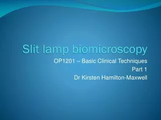
BasicsDefinition and Applications Definition The purpose of a slit lamp is the biomicroscopy of the patient‘s eye under different lighting conditions. The slit lamp projects a bright and homogenously illuminated slit onto the eye which is variable in length, width, angle and light intensity. Fields of Application The primary field of application is the anterior eye segment (cornea, anterior chamber, lens, anterior vitreous). Using additional optics enables the user to also examine the posterior eye segment as well as the anterior chamber angle.
BasicsDesign Principles • components: microscope, slit projector, instrument base • carrier arms for microscope and slit projector can be swiveled around a common axis • swivelling axis is located in the focal plane of microscope and slit projector
BasicsDesign Principles - Biomicroscope • Zeiss slit lamps:Galilei type microscope • common front objective • parallel beam path • 3 or 5 magnification steps • other manufacturers also offer Greenough type microscopes • two separate, tilted beam paths • only 2 magnification steps • very few accessories
BasicsDesign Principles - Slit Projector • purpose: to project a slit image focused on the patient‘s eye • the slit image is variable in length, width and angle • light source: usually halogen (high color temperature) • filters: blue, green (redfree), diffusor, heat absorbing filter
BasicsDesign Principles - Instrument Base • functional coupling of carrier arms of microscope and slit projector • slit projector and stereo microscope can independantly be swiveled around a common axis • Axis is located below patient’s eye • both slit image and observation are in focus in the axial plane • three-dimensional positioning by joystick
Basic Functions of the Slit LampSlit Width narrow slit circle shaped, if fully opened slit width is adjusted continuously
Basic Functions of the Slit LampSlit Length short slit long slit slit length is adjusted in steps and continuously
Basic Functions of the Slit LampSlit Rotation vertical slit horizontal slit vertical slit slit rotation can be adjusted continuously by ±90°
Basic Functions of the Slit LampSlit Decentration decentered slit slit can be decentered continuously by ±4°
Basic Functions of the Slit LampTilting Prism angle of incidence 0° angle of incidence 20° tilting prism can be tilted by 0° to 20° continuously positions 0°, 5°, 10°, 15°, 20° indexed
Types of Illumination Forms of direct Illumination
Types of IlluminationDirect Diffuse Illumination Principle • illumination of the eye with a broad, unfocused light beam • usage of diffusor • microscope positioned at 0° • magnification 5x ... 12x Applications • Overview • general assessment of anterior eye, eye lids • assessment of contact lenses
Types of IlluminationDirect Diffuse Illumination IOL iris supported anterior chamber lens in diffuse illumination Bildquelle: Universitäts-Augenklinik Jena
Types of IlluminationDirect Focal Illumination - Optic Section Principle • Illumination and observation are focused in the same plane • slit width ca. 0,1 to 0,3mm Applications • mainly findings in the cornea and lens • opacities, scars, vessels • good perception of the depth of findings
Types of IlluminationDirect Focal Illumination - Optic Section Cataract anterior cortical opacities, nucleosclerosis and posterior opacities Bildquelle: www.atlasophthalmology.com
Types of IlluminationDirect Focal Illumination - Optic Disc Principle • Illumination and observation are focused in the same plane • slit width ca. 2 to 4mm Applications • mainly findings in the cornea and lens • opacities, scars, vessels • good perception of shape and size of findings
Types of IlluminationDirect Focal Illumination - Optic Disc Cyst on Pupillary Edge Cyst on pupillary edge stems from usage of too strong miotica Bildquelle: www.atlasophthalmology.com
Types of IlluminationDirect Focal Illumination - Conical Beam Principle • assessment of particles floating in the anterior chamber by illuminating with a light beam • Tyndall‘s phenomenon • pinpoint illumination 0,3 - 0,5mm Applications • assessment of particles in aqueaous humor • inflammation cells, pigmented cells, metabolic waste
Types of IlluminationDirect Focal Illumination - Conical Beam cells in anterior chamber cells in anterior chamber as a sign of uveitis Bildquelle: www.atlasophthalmology.com
Types of IlluminationTangential Illumination Principle • a narrow light beam is projected almost parallel along the structure to be observed • elevated structures are visible by shadowing Applications • elevated abnormities or changes in the iris • tumors, cysts
Types of IlluminationTangential Illumination Iris Iris in tangential illumination
α α
α α 0° Types of IlluminationSpecular Illumination Principle • angle of incidence = angle of reflection • observation and illumination have same angle to perpendicular axis • slit width < 4mm Applications • assessment of surfaces • assessment of tear film • endothelial cell layer
α α 0° Types of IlluminationSpecular Illumination endothelial cells endothelial cell layer magnified ca. 192x Bildquelle: Carl Zeiss Meditec
Types of Illumination Forms of indirect Illumination
Types of IlluminationIndirect focal Illumination Principle • illumination by stray light • slit is slightly decentered so that stray light is created in direct neighbourhood of the finding • slit width ca. 2 to 4mm Applications • mainly corneal lesions and scars
Types of IlluminationDirect Retro-Illumination from the Iris Principle • Illumination of the finding with indirect light rflected from the iris • observation with light background • medium slit width, ca. 2 to 4mm Applications • Infiltrations, small scars, corneal vessels, micro cysts, vacuoles • with this illumination findings are made visible with high contrast
Types of IlluminationDirect Retro-Illumination from the Iris Keratitis Superficialis Punctata finding after moderate cauterization by acid, defects of epithelium and conjunctiva have been stained with bengal rose Bildquelle: www.atlasophthalmology.com
Types of IlluminationIndirect Retro-Illumination from the Iris Principle • Illumination of the finding with indirect light reflected from the iris • observation with dark background • medium slit width, ca. 2 to 4mm Applications • Infiltrations, small scars, corneal vessels, micro cysts, vacuoles
Types of IlluminationIndirect Retro-Illumination from the Iris Keratitis Punctata, contact lens wearer multiple erosions of the central cornea due to inappropriate contact lens fitting Bildquelle: www.atlasophthalmology.com
Types of IlluminationRetro-Illumination from the Lens Principle • Illumination of the finding with indirect light reflected from the lens • observation with light background • medium slit width, ca. 2 to 4mm Applications • corneal defects, foreign bodies, scars • (type of illumination not frequently used)
Types of IlluminationRetro-Illumination from the Lens no example
Types of IlluminationRetro-Illumination from the Fundus Principle • Illumination of the finding with indirect light reflected from the fundus • observation with red/yellowish background • dilated pupil Applications • abnormities in the anterior vitreous, lens, anterior chamber, cornea • findings are visible like silhouettes
Types of IlluminationRetro-Illumination from the Fundus Aniridia missing iris and zonular cataract made visible by retro-ilumination Bildquelle: www.atlasophthalmology.com
Types of IlluminationIris-Transillumination Principle • transillumination of the iris by indirect light reflected from the fundus • half dilated pupil (3 to 4mm) • Illumination and observation at ca. 0° Applications • Visualization of defects of the pigment layer of the iris
Types of IlluminationIris-Transillumination Albinism Iris-Transillumination shows the light transmission of the iris Bildquelle: www.atlasophthalmology.com
Types of IlluminationSclerotic Scatters Principle • Illumination of the limbus region with a broad light beam at an angle of 45° - 60°, decentered slit • total reflection of the incoming light at inner corneal boundaries (endothelium and epithelium) Applications • scars, foreign bodies, corneal defects • irregularities in the cornea cause straylight
Types of IlluminationSclerotic Scatters corneal scar corneal scarring after infection Bildquelle: www.atlasophthalmology.com
Fundus Observation and GonioscopyContact Glasses Contact Glasses • additional tool for fundus observation with the slit lamp • mostly direct: erect and non mirrored image of the fundus • required: dilated pupil, use of gliding liquid Fundus Image Microscope Bildquelle: www.ocular-instruments.com
Inverted fundus image microscope Fundus Observation and GonioscopyLenses Lenses • additional tool for fundus observation with the slit lamp • mostly indirect: upside-down and mirrored image of the fundus (convex optics) • non contact • required: dilated pupil Bildquelle: www.ocular-instruments.com
Fundus Observation and GonioscopyGonioscopy Three mirror contact glass • Goldmann contact glass • central lens: posterior pole • 73° mirror: equator • 67° mirror: ora serrata • 59° mirror: anterior chamber angle central lens Bildquelle: www.ocular-instruments.com
73° mirror Fundus Observation and GonioscopyGonioscopy Three mirror contact glass • Goldmann contact glass • central lens: posterior pole • 73° mirror: equator • 67° mirror: ora serrata • 59° mirror: anterior chamber angle Bildquelle: www.ocular-instruments.com
67° mirror Fundus Observation and GonioscopyGonioscopy Three mirror contact glass • Goldmann contact glass • central lens: posterior pole • 73° mirror: equator • 67° mirror: ora serrata • 59° mirror: anterior chamber angle Bildquelle: www.ocular-instruments.com
59° mirror Fundus Observation and GonioscopyGonioscopy Three mirror contact glass • Goldmann contact glass • central lens: posterior pole • 73° mirror: equator • 67° mirror: ora serrata • 59° mirror: anterior chamber angle Bildquelle: www.ocular-instruments.com
Fundus Observation and GonioscopyExample: Fundus retinal scar microscope Bildquelle: UAK Jena / Carl Zeiss
Fundus Observation and GonioscopyExample: Anterior Chamber Angle blood in chamber angle vessels in chamber angle