Download
1 / 70
810 likes | 1.3k Views
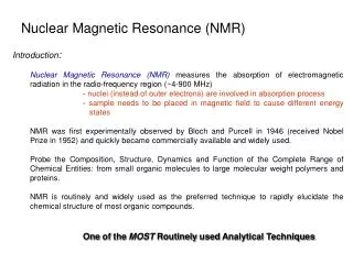
NMR = Nuclear Magnetic Resonance NMR spectroscopy is the most powerful technique available to organic chemists for determining molecular structures. Looks at nuclei with odd mass numbers or odd number of protons: 1 H, 13 C , 15 N, 19 F, 31 P.

E N D
NMR = Nuclear Magnetic Resonance NMR spectroscopy is the most powerful technique available to organic chemists for determining molecular structures. Looks at nuclei with odd mass numbers or odd number of protons: 1H, 13C, 15N, 19F, 31P
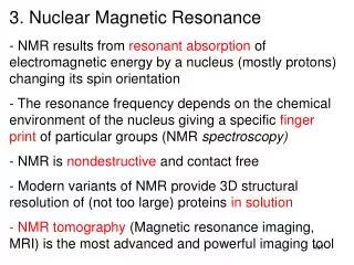
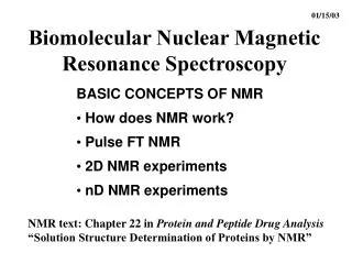
NMR spectroscopy that is looking at 1H nuclei is called proton NMR or 1H-NMR. If you just say NMR, it will generally be assumed you’re talking about proton NMR. NMR looking at 13C nuclei is called 13C-NMR. Nuclei with odd mass numbers or number of protons have nuclear spin states. Spinning nuclei generate magnetic fields.
Spinning nuclei will line up with or against an external magnetic field (B0). Aligned against the field Aligned with the field
Alignment with the field (lower energy) is called the spin state. Alignment against the field (higher energy) is called the spin state. The difference in energy between and : E. In the absence of an external magnetic field, orientations are random. Application of the external field forces nuclei into the or spin states. B0 E no external field
A photon of the correct energy (E) can cause flipping from the spin state to the spin state. When flipping occurs, the energy of that photon is absorbed. When the combination of the external magnetic field strength and photon energy produce flipping, the nucleus is “in resonance” with the magnetic field and that frequency of electromagnetic radiation. E is proportional to the external magnetic field strength. h E = B0 2 where B0 is measured in gauss and is the gyromagnetic ratio – unique for each kind of nucleus
h E = B0 2 For a proton, = 26,753 sec1 gauss1 Since E = h h h = B0 2 Factoring out h, gives 1 = B0 2 The frequency () and field strength (B0 ) are directly proportional.
1 = B0 2 This equation tells us that for a frequency of 60 MHz (radio frequency or RF), a magnet with a strength of 14,092 gauss is needed for resonance with a bare proton (H nucleus). A radio frequency of 300 MHz requires a magnet 5x stronger (70,459 gauss) for resonance with a bare proton.
But nuclei in molecules are not bare, isolated nuclei – they are surrounded by clouds of electrons. In the presence of an external magnetic field, an electron cloud has an induced magnetic field that opposes the external field. This means it takes a stronger external field to get resonance – a stronger field is needed because the electron cloud effectively shields the nucleus. The effective magnetic field experienced by the nucleus is the strength of the external field minus the shielding effect. Beffective = Bexternal Bshielding
The electron cloud density (and thus shielding) is different for different nuclei due to different chemical environments (magnetic environments). less shielded proton due to electron withdrawing effect of electronegative oxygen requiring lower field strength to get resonance at a specific more shielded protons requiring higher field strength to get resonance at a specific The differences in shielding mean that the we can detect the differences in the chemical environments of the nuclei in an NMR spectrometer.
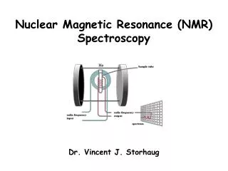
NMR spectrometer: RF is held constant – common frequencies are 60 MHz, 100 MHz, and 300 MHz. Magnetic strength is varied
upfield downfield increasing magnetic field strength
Chemical shift Chemical shift for protons = the difference between magnetic field strength needed for resonance of a given proton and the magnetic field strength needed for resonance of a proton in tetramethylsilane (TMS). Due to the low electronegativity of the central silicon atom, protons in TMS are more shielded than any other protons likely to show up in organic compounds. Proton signals in most organic compounds will therefore be shifted downfield from TMS (i.e. require weaker fields for resonance).
The amount of downfield shifting from TMS is expressed in Hz and is divided by the radio frequency of the instrument. (Even though NMR spectrometers vary magnet strength, remember that frequency is proportional to magnetic field strength.) chemical shift = shift downfield from TMS in Hz instrument frequency in MHz Since Hz units cancel, the chemical shift is unitless. Since MHz are 1,000,000 times Hz, the amount of chemical shift is called parts per million (ppm). The chemical shift is symbolized by (delta scale).
The advantage of expressing chemical shift data on the delta scale is that it is easily allows for the comparison of data from instruments that are operating at different field strengths.
The higher field strength instruments are used because they give higher resolution and higher sensitivity. The main drawbacks to the higher field strength instruments are initial cost and very high maintenance cost.
300 MHz NMR spectrum of methanol: Ultimately, the number of signals indicates the number of different chemical environments in the molecule.
Since the number of signals is related to how many different chemical environments are present in a molecule, it is useful to be able to recognize groups of chemically equivalent protons. How many groups of chemically equivalent protons are present in each of the following compounds. Hint: Think about symmetry. If different chemical shifts are so close that the signals cannot be resolved, the protons are called accidentally equivalent. Accidental equivalence can only be determined experimentally.
Diastereotopic protons are in different chemical environments (i.e. are not chemically equivalent) and therefore give different chemical shifts (may only resolve in 300 MHz instrument). Diastereotopic protons are identified by seeing if you get diastereomers by replacing them. Are these protons chemically equivalent? imagine replacing each with Br or some other atom What is the relationship between these two structures?
Are these protons chemically equivalent? imaginary replacement R enantiomer S enantiomer Replacement did not produce diastereomers, therefore the chemical environments are the same. Therefore both protons will give the same chemical shift. The protons in question are enantiotopic protons which are chemically equivalent. NMR can not distinguish enantiomer.
How many groups of chemically equivalent protons are present in the following compounds?
Again, different protons have different expected chemical shifts – depending upon their different chemical environments. CH3-CH2-CH2-CH3 CH3-CH2-CH2-CH2-Br = 0.9 1.3 1.3 0.9 0.9 1.3 1.7 3.4 deshielding caused by nearby Br = 0.0 0.0 0.4 2.5
Predict the expected chemical shifts of the following groups of chemically equivalent protons. Chemical shift values are approximately additive – when you predict chemical shifts you need to take into account additive effects.
Groups on a benzene ring can cause downfield or upfield shifts depending upon whether the group is electron withdrawing or electron donating.
Origins of chemical shifts In some cases, electron withdrawing effects cause deshielding (and electron donation causes more shielding). Ex. CH3-CH2-CHO CH3-CH2-NO2 In other cases, shielding and deshielding are caused by magnetic effects of nearby pisystems…
Aldehyde protons are strongly deshielded by a combination of electron withdrawing and magnetic effects. = 9 - 10
Carboxylic acid protons are very strongly deshielded due to being attached to an O which is attached to an electron-withdrawing C=O. ( = 10 – 12) What else can you say about the compound that gave the above spectrum?
Peak area – related to number of protons in a given chemical environment. Indicated by an “integral trace” on NMR spectrum. (height of trace is proportional to peak area) (peaks with bigger areas are usually taller) a b c a a 3.0 d 6 spaces total b 1.5 c 1.0 d 0.5 Ratio of protons - a:b:c:d = 3.0 : 1.5 : 1.0 : 0.5 To get whole numbers multiply by 2 to get: 6 : 3 : 2 : 1
a b c a a 3.0 d 6 spaces total b 1.5 c 1.0 d 0.5 A ratio of 6 : 3 : 2 : 1 could represent a compound with a total of 12 H’s (6 + 3 + 2 + 1 = 12) or… Any compound with the same ratio and a multiple of 12 H’s Ex. 12 : 6 : 4 : 2 ratio for a total of 24 H’s
a b c a a 3.0 d 6 spaces total b 1.5 c 1.0 d 0.5 Or if total number of H’s is known to start with, figure out how many spaces = 1 H (You have a molecular formula.) 6 spaces = 12 H’s means 0.5 spaces = 1 H The integral with 3 spaces then represents 6 H’s giving that signal.
a b c a a 3.0 d 6 spaces total b 1.5 c 1.0 d 0.5 Integral traces are not always this neat and well-defined. Sometimes deciding where traces start and end is tricky – may require some trial and error to get ratios that make sense.
Insert NMR with integration curve and have class determine ratio on a handout.
Spin-Spin Splitting Signals of protons often do not show up as single peaks – their signal can be split by magnetically coupled protons. Because a proton we’re interested in may be near other protons, the magnetic fields of those nearby protons can reinforce (add to) or oppose (subtract from) the external magnetic field – shifting a signal downfield or upfield. Downfield shift due to one adjacent proton adding to field strength Upfield shift due to one adjacent proton subtracting from field strength Expected signal location
The more magnetically coupled protons there are, the more complex splitting gets because all adjacent protons could be spinning in the same direction, all adding or all opposing the external field or some could be spinning in opposite directions giving mixed effects. Ultimately, this splitting will tell you the number of protons on adjacent carbons. Notice that the peak area ratios reflect the number of combinations that cause each peak
The splitting pattern ultimately follows an N + 1 Rule: N equivalent coupled protons split a signal into N + 1 peaks. Relative peak areas Number of peaks (multiplicity) N 0 1 (singlet) 1 1 2 (doublet) 1 : 1 2 3 (triplet) 1 : 2 : 1 3 4 (quartet) 1 : 3 : 3 : 1 4 5 (quintet) 1 : 4 : 6 : 4 : 1 5 6 (sextet) 1 : 5 : 10 : 10 : 5 : 1 6 7 (septet)
H H 1 4 CCC 3 2 Most splitting is caused by adjacent protons. Ph-CH2-CH3 Some splitting is caused by protons on the same carbon (only if diastereotopic). Splitting by protons separated by a total of 4 or more bonds is normally not observed (but there are exceptions) These protons should split each other’s signals – splitting is reciprocal. These protons will split each other’s signal Too far away to be magnetically coupled
Characteristic splitting patterns to know: Peaks representing magnetically coupled protons often “lean” toward one another. • Pattern for isopropyl group • Pattern for p-disubstituted benzene (1 withdrawing group, 1 donating group)
Coupling constants (J) – how much proton signals split each other. The magnitude of the J value is also reciprocal.
Areas of the NMR Spectra can be expanded to show greater detail. Below it is easier to measure the value of thecoupling constant in the expanded view.
Assign the proton signals in the following spectrum of 4,4-dimethylcyclohe-2-en-1-one.
Complex splitting When a signal is split by two or more protons that have different coupling constants, the pattern of the peaks no longer follows an N + 1 rule.
a c Protons a and b are trans with coupling constant (Jab) = 17 Hz b Protons a and c are cis with Jac = 11 Hz You can draw a splitting tree to show the expected pattern of peaks for the signal of proton a – starting with the biggest split.
a c b Protons a and b are trans with coupling constant (Jab) = 17 Hz Protons b and c are geminal with Jbc = 1.4 Hz
Label each set of peaks in the NMR spectrum with the letter of the corresponding protons in the following structure:
Label the groups of protons in the structure below with the letter corresponding to the peak in the NMR spectrum associated with those protons: d e a b c
Determine the structure for a compound with a formula of C9H10O2 if an IR spectrum shows a strong peak at 1705 cm1.
Determine the structure for a compound with a formula of C9H10O2 if an IR spectrum shows a strong peak at 1735 cm1.