Download
1 / 49
490 likes | 640 Views
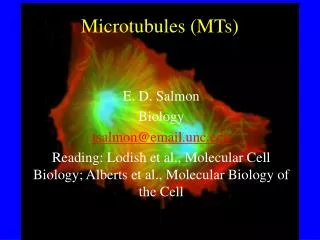
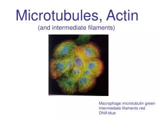
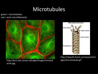
Microtubules, Motors and Membranes. Transport on Microtubules. In neurons there is visible transport of vesicles from cell body to growth cone Transcription and translation and membrane biosynthesis in cell body Need to get material to growth cone to elongate Axonal transport

E N D
Transport on Microtubules • In neurons there is visible transport of vesicles from cell body to growth cone • Transcription and translation and membrane biosynthesis in cell body • Need to get material to growth cone to elongate • Axonal transport • Fast anterograde (3µm/sec)-vesicles • Intermediate anterograde (0.6µm/sec)-mitochondria • Slow anterograde- (0.002-0.03µm/sec) -proteins • Retrograde- 2µm/sec
Giant Squid axoplasm can be extruded and watched under microscope • Can watch vesicles move on mt’s • Now use brain mt’s and squid axoplasm • vesicles move with ATP added (2µm/sec) • vesicles bind but don’t move with AMPPNP • now isolate proteins that bind to mt’s in the presence of AMPPNP but elute with ATP! • Kinesin is discovered
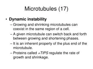
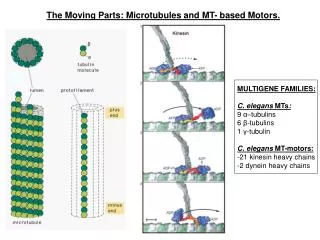
Kinesins • Moves toward the (+) end (1-2 µm/sec) • 2 x 124kd + 64kd complex • Double headed ATP motor with a tail that binds cargo • 4 families involved in vesicle movement • 3 families involved in spindle function • Some are actually (-) end directed
Dynein • Huge protein complex • 2-3 500 kd proteins • several intermediate and light chains • dynactin complex • 4 proteins including an actin-related protein (ARP) • regulates dynein? • (-) end directed ATPase motor (1-14µm/sec) • Three classes of cytoplasmic plus flagellar • One looks vesicular • One is near Golgi • One is in punctate structures of unknown origin
Microtubule motors in vitro 1- Put motor on slide and add microtubules- the motor pushes the microtubule along 2- add vesicles to microtubules on slide and the vesicles are moved on the microtubules
Terasaki et al. • DiOC6 stains mitochondria +ER • Shows a reticular network in cells • Co-localizes with mt’s • Depolymerize mt’s and it collapses, but slower than mt’s • During regrowth, the ER follows the mt’s
Dabora and Sheetz • Make an membrane prep from CEF cells • Add to mt’s on coverslip • the vesicles are pulled out into a reticular network • requires ATP • inhibited by AMPPNP and vanadate (requires kinesin and dynein motors) • looks like Terasaki’s ER • Recent- Kinesin binding protein found on cytoplasmic face of ER- Kinectin
Turner and Tartakoff • Depolymerize mt’s with nocodazole • Golgi vesiculates • required energy • Now remove nocodazole • mt’s reform • Golgi coalescence • requires energy
Golgi (green) Microtubules’s (red) Add nocodazole
ER-Golgi- Transport • ER to Golgi traffic visualized in living cells John F. Presley, Nelson B. Cole, Trina A. Schroer, Koret Hirschberg, Kristien Zaal and Jennifer Lippincott-SchwartzNature,Volume 389, Pages 81-85, 1997 • Kinetic Analysis of Secretory Protein Traffic and Characterization of Golgi to Plasma Membrane Transport Intermediates in Living Cells J. Cell Biol., Volume 143, Number 6, 1998 1485-1503 Koret Hirschberg, Chad M. Miller, Jan Ellenberg, John F. Presley, Eric D. Siggia, Robert D. Phair,§ and Jennifer Lippincott-Schwartz • Use GFP probes to visualize ER to Golgi transport • VSVG ts mutant- doesn’t fold correctly at low temperature • 40°C- blocked in ER • 15°C- blocked in pre-Golgi • 32°C- transports normally
Hirschberg et al. Cells incubated at 40C overnight to block in ER and then released to visualize dynamics of transport
Presley et al. Cells held at 40 to block in ER and then released to see movement to Golgi
Presley et al. Cells blocked in pre-golgi at 15 then released to 32 to see movement to Golgi
Presley et al. Movement of pre-golgi structures (15C) in the presence of nocodazole
Presley et al. Overexpress Dynamitin at 15C and then shift to 32 (release to Golgi)
Mitochondria and Microtubules • Mitochondria also coalign with Mt’s in cell • They are elongated into tubules along the length of mT’s • Recently, a kinesin homolog (Kif1B) has been found to be specific for mitochondria
Lysosomes and Microtubules • Label endosomal system see extensive network of vesicles and tubules clustered around MTOC • Nocodazole causes dispersal • Movements of individual vesicles ceases when mt’s depolymerized • repolymerize mt’s and they recluster at MTOC
Melanophores • Vesicles move bidirectionally on Mt’s to change the color of cells in fish scales • Melatonin stimulating hormone causes vesicles to disperse • cAMP increases • Melatonin causes them to aggregate near the nucleus • cAMP decreases • Dynein does inward movement • Kinesin + Myosin V does outward
Models for bidirectional vesicle transport (Gross et al. The Journal of Cell Biology, Volume 156, Number 4, February 28, 2002 715–724)
Melanophore papers • Movements of melanophores in cells • Functional Coordination of Microtubule and Actin Based Motility in Melanophores. V. I. Rodionov, A. J. Hope, T. M. Svitkina and G.G. Borisy Curr. Biol., 8(3): 165-168, 1998
Movements are microtubule dependentGross et al. The Journal of Cell Biology, Volume 156, Number 5, March 4, 2002 855–865
All three motors stay bound to melanosomes during movement(Gross et al. The Journal of Cell Biology, Volume 156, Number 5, March 4, 2002 855–865
Dynactin complex(Deacon et al. The Journal of Cell Biology, Volume 160, Number 3, February 3, 2003 297–301) • Complex that binds dynein to cargo • Inhibition of function by overexpressing dynamitin ( a component of dynactin complex) inhibits both aggregation and dispersal • Found dynamitin binds to Kinesin as well as dynein • Thus the dynactin complex may regulate both with some kind of bidirectional on/off switch
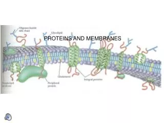
Vesicle trafficking: Golgi-TGN-sorting in epithelial cells • Problem: All secretory, lysosomal, membrane proteins sort from ER to Golgi and then are distributed in the TGN to different sites • How is the sorting accomplished? • In Epithelial cells you sort cargo to the apical and basolateral membranes. How?
Use fluorescent labeled markers that sort to apical (CFP) or basolateral (YFP) part of the cell. Keller et al. Nature Cell Biology, 3:140-147 2001 • Transfect in probes • Block transport at low temperature • Release block and image • Most probe is initially in the same vesicle (yellow) • Over time, they sort into red and green vesicles • Can see tubules pull out of a single sorting compartment that are either red or green • Mechanism unknown
Movements near the membrane are not diffusion! (Keller et al.) TIRF imaging of basolateral side of cell Red- endocytic marker (dextran uptake) Green- basolateral secretory marker
Membrane Tension • Membranes seem to be under tension-note the pulling out of membranes along microtubule’s in vitro looks like ER • Sorting seems to involve stretching of the sorting compartment membranes into tubules • There is now evidence that in the TGN, you don’t bud off small vesicles, but large interconnected networks of tubules and vesicles • Brefeldin A Blinkout • Brefeldin A blocks the transport from ER to Golgi • Does not block recycling from Golgi to ER • Add bfa and watch the Golgi disperse- it is not gradual, it is explosive!!
Future? • Challenge is to figure out the specific function of each motor • Where is it? • What is it’s cargo? • What turns the motor off and on? • Organization of Golgi, ER, lysosomes by motors and how function is interrelated