Download
1 / 38
380 likes | 660 Views
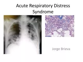
ARDS Acute Respiratory Distress Syndrome NURS 504 Liberty University Jenny Holloway June 10, 2012 Dr. Rainey. ARDS : wet lung , white lung acute lung injury characterized by coughing and rales ; inflammation of the lungs which become stiff and fibrous and cannot exchange oxygen

E N D
ARDS Acute Respiratory Distress Syndrome NURS 504 Liberty University Jenny Holloway June 10, 2012 Dr. Rainey
ARDS: wet lung, white lung acute lung injury characterized by coughing and rales; inflammation of the lungs which become stiff and fibrous and cannot exchange oxygen (NCCBLED, 2009)
ARDS PATHOPHSIOLOGY • Inflammatory cells and proteinaceous fluid accumulate in the alveolar spaces leading to a decrease in diffusing capacity and hypoxemia.
Acute lung injury leads to: • An unregulated systemic inflammatory response lead to damaged capillary membranes. These membranes leak, further damaging the alveolar membrane. • Fluid enters the alveoli resulting in significant tissue hypoxemia leading to acidosis
Predisposing Conditions that can lead to ARDS • Sepsis (#1 cause) • Severe Pneumonia • Aspiration • Near Drowning • Smoke inhalation • Multiple blood product transfusions • Trauma • DIC • Drug overdose: Heroin, methamphetamine, cocaine • Acute pancreatitis • Severe Burns
THREE PHASES OF ARDS • PHASE I (Exudative) • Acute lung injury damage leaking fluid excessive fluid in the lungs • Arterial oxygenation poor low PaO2 • Some patients recover during this phase, most progress to phase II • Lasts about a week if recovery does not occur
THREE PHASES OF ARDS • PHASE II (Fibroproliferative) • Connective tissue proliferates (or reproduces rapidly) enlarged air spaces and fibrosis tissue • Most clients who die during this phase die of Multiple Organ failure • Lasts 3-10 weeks
THREE PHASES OF ARDS • PHASE III • Resolution of inflammation • O2 improves and weaning of ventilatory support can begin • Lasts 6-12 months • Lasting damage will differ from client to client
A diffuse inflammatory process • Circulating neutrophils are activated and become “sticky.” They adhere to the vascular endothelium and spill their cytoplasmic granules which then damage the endothelium leading to leaky capillaries. The result: An exudative fluid accumulates in the lung parenchyma, which leads to further damage locally (i.e. alveolar cell damage) decreasing oxygenation and lung compliance. • Fibrin Deposition. Fibrin release is triggered by tissue factor. Over time the fibrin can later remodel to form fibrosis. Marino, P.L. The ICU Book. 3rd Ed. Lippincott Williams & Wilkins. Philadelphia. 2007
Pulmonary Edema CARDIAC RELATED PULMONARY EDEMA NON-CARDIAC PUMONARY EDEMA Infiltrates are more homogeneous No pleural effusions No Kerley B’s Radiographic evidence lags behind clinical signs and symptoms (i.e. the CXR is unimpressive given the degree of hypoxemia • Patchy infiltrates appearing in the lung bases first • Effusions may be present • Clinical signs and symptoms lag behind radiographic evidence (i.e. CXR is more impressive than the degree of hypoxemia)
PULMONARY EDEMA Cardiac related Non-cardiac related http://www.homeofpoi.com/lessons_all/teach/Firebreathers-Lung-or-ARDS-11_52_198
Cardiac related Pulmonary Edema • Vascular Endothelium breaks under stress easily, however it also repairs itself quickly • Cardiogenic edema often develops quickly and can resolve quickly because vascular endothelium is able to repair itself quickly
NON-CARDIAC PULMONARY EDEMA • Alveolar epithelium is quite resistant to damage. It withstands greater force before becoming damaged. However, once “broken” it takes much longer to heal than endothelium. • Cellular damage in Non-Cardiogenic edema runs along a spectrum from predominately vascular endothelial damage to predominately alveolar epithelial damage
VENTILATION Negative Pressure Positive Pressure
Positive-pressure Ventilators • Modes with positive-pressure ventilators • Continuous positive airway pressure (CPAP) • Bilevel ventilator (BiPAP) • Assist-control mode ventilation (ACMV) • Synchronized intermittent mandatory ventilation (SIMV) • Positive end-expiratory pressure (PEEP) • Pressure-support ventilation (PSV) • Pressure-control ventilation (PCV)
Ventilatory Management • Low tidal volume mechanical ventilation • In ARDS there is a large amount of poorly compliant (i.e. non-ventilating) lung and a small amount of healthy, compliant lung tissue. Large tidal volume ventilation can lead to over-inflation of the healthy lung tissue resulting in ventilator-induced lung injury of that healthy tissue. • PEEP • Setting a PEEP prevents further lung injury due to shear forces by keeping airways patent during expiration
Ventilatory Management • Is there such thing as too low a TV? • Tidal Volume must be sufficient for gas exchange to take place. Permissive hypercapnia is the term used to state that a certain degree of hypercapnia and its resulting acidemia can be allowed in order to maintain lung-protective TVs. • Absolute limits is unclear, but a pH of 7.2-7.25 and a PCO2 of 60-70 mm Hg is a good cut off range. • Is there such thing as too much PEEP? • PEEP serves to help open less compliant alveoli and keep alveolar open during expiration, but it too can lead to overinflation of alveoli that are already maintaining aeration. • Setting PEEP too high also increases intrathoracic pressure leading to decreased venous return. • Start patients at a PEEP trial of 5 – 12 cm H2O and increase if needed.
PHARMACOLOGIC MANAGEMENT OF ARDS • Fluid for hemodynamic management • Diuretics • Beta-agonist • Inhaled nitric oxide • Steroids • NSAIDS • Remember, the #1 goal in therapy is to decrease tissue ischemia. We must maintain ARDS patient’s CO (cardiac output) to insure tissue profusion.
FLUIDS • Crystalloid • Isotonic • 0.9% • Lactated Ringers (LR) • Action/Use • No fluid shift • Vascular expansion • Electrolyte replacement • Nursing Considerations • May cause fluid overload • Generalized edema • Dilutes hemoglobin • May cause electrolyte imbalances • Proinflammatory in large doses
TO DIURESE OR TO NOT DIURESE YES NO Diuretics are not anti-inflammatory agents: lung infiltrates in ARDS are neutrophils and proteins, NOT edema Hemodynamic compromise: tissue oxygenation #1 concern. Aggressive diuretics decrease venous pressures leading to decrease CO and increased tissue ischemia • Diuretics have been shown to decrease any pulmonary edema that is present, increase lung compliance, and improve gas exchange. However they have shown no survival benefit.
BETA-AGONIST • Short acting: • Albuterol • 2-4mg tid • 1-2 inhalations q4-6hrs • May be given by nebulizer • Xopenex • 0.63mg tid q 6-8 hrs by nebulizer • May increase to 1.25mg tid if no response • Action/Use • Treat and prevent bronchospams • Nursing Considerations • May cause tachycardia • May cause nervousness, anxiety • May cause cardiac arrhythmias
INHALED NITRIC OXIDE • Selective pulmonary vasodilator • Given as inhaled gas • Action/Uses • No systemic hemodynamic effects • Improves Oxygenation • Decrease Pa pressures reducing right ventricular afterload • Nursing Considerations • No study demonstrates a reduction in mortality
STEROIDS • Corticosteroids • Methyl-prednisone • Imflammation • 10-80mg IV daily • Action/Use • Stablizeslysosomalmembrance and prevents the release of proteolyticenzmes during inflammatory process • Decreases the production of lymphocytes and eosinophils, blocks the release of cytokines • Nursing Considerations • Monitor for s/s of cushing’s syndrome • Monitor glucose levels • Monitor serum potassium
NSAIDS • Specifically Ibuprofen • 400mg q 4-6 hrs; maximum daily dose 3.2 grams • Actions/Uses • Anti-inflammatory agent, fever • Nursing Considerations • May cause GI upset • May cause GI bleeding • Monitor renal function
HIGH MORTALITY RATE • Despite early intervention mortality rate 31-74% • Main cause of death is usually not respiratory related • Die with ARDS not from ARDS • Respiratory failure is the cause of death in 9-16%
NURSING DIAGNOSIS • Risk for Acute Confusion • Ineffective Airway Clearance • Ineffective Breathing Pattern • Impaired Gas Exchange • Decreased Cardiac output • Dysfunctional Ventilatory Weaning Response • Carpenito-Moyet, L. (2006). Handbook of nursing diagnosis. (11th ed.). Philadelphia: PA, Lippincott.
NURSING DIAGNOSIS • Risk for Imbalanced Fluid Volume • Imbalanced Nutrition: Less Than Body Requirements • Risk for Infection • Acute Pain • Anxiety • Carpenito-Moyet, L. (2006). Handbook of nursing diagnosis. (11th ed.). Philadelphia: PA, Lippincott.
GOALS • Client will • Be oriented with each interaction • Receive adequate ventilatory support • Be free of pulmonary tissue damage • Maintain patent airways • Maintain adequate cardiac output • Receive adequate nutrition • Be free of signs, symptoms of infection • Carpenito-Moyet, L. (2006). Handbook of nursing diagnosis. (11th ed.). Philadelphia: PA, Lippincott.
GOALS • Client will • Have no development of thrombosis • Manage pain successfully • Cope with or be free from anxiety • Carpenito-Moyet, L. (2006). Handbook of nursing diagnosis. (11th ed.). Philadelphia: PA, Lippincott.
NURSING INTERVENTIONS • Common interventions • Lab work and specimens • Monitor • Vital signs hourly • Oxygenation status • Neurologic status • Lung and heart sounds • Provide analgesia, anxiolytics, sedation • Beta-agonist to maintain patent airways • Maintain HOB at 30° or higher • Carpenito-Moyet, L. (2006). Handbook of nursing diagnosis. (11th ed.). Philadelphia: PA, Lippincott.
NURSING INTERVENTIONS • Prone position as tolerated 3–4x/day • YES PRONE • Suction airways as needed • Monitor hemodynamic status • Monitor renal function • Place Foley catheter • IV fluids as needed • Monitor glucose levels • Assess peripheral pulses • Carpenito-Moyet, L. (2006). Handbook of nursing diagnosis. (11th ed.). Philadelphia: PA, Lippincott.
EVALUATION • Client maintains oxygen saturation greater than 90% • Vital signs remain within acceptable limits • Client’s airway remains clear • ABG results indicate acid-base balance maintained • Carpenito-Moyet, L. (2006). Handbook of nursing diagnosis. (11th ed.). Philadelphia: PA, Lippincott.
References • Bhadade, R. R., A., Harde, M. J., & Khot, A. A. (2011). Clinical characteristics and outcomes of patients with acute lung injury and ARDS. Journal Of Postgraduate Medicine, 57(4), 286-290. • Carpenito-Moyet, L. (2006). Handbook of nursing diagnosis. (11th ed.). Philadelphia: PA, Lippincott. • Edmunds, M. W. (2009). Pharmacology for the primary care provider. (3rd ed.) St Louis, MO: Mosby. • Lehne, R. A. (2007). Pharmacology for nursing care. St Louis, MO: Saunders. • Marino, P.L. (2007). The ICU Book. (3rded). Philadelphia: Pa, Lippincott Williams & Wilkins • North Carolina Concept Based Learning Education Board (2011). Nursing : A Concept Based Approach to Learning. Upper Saddle River, NJ: Pearson. • Tang, B.M.; Craig, J., Eslick, G., Seppelt, Il, & McLean, A. (2009). Use of corticosteroids in acute lung injury and acute respiratory distress syndrome: A systematic review and meta-analysis. Critical Care Medicine, 37(5), 1594-1603. • Taylor, M. (2005). ARDS diagnosis and management implications for the critical care nurse. Dimensions of Critical Care Nursing, 24(5), 197-206.
References • Turkoski, B. Drug information handbook for advanced practice nursing. (10th ed.). Hudson, OH: Lexi-Comp. • Villar, J. (2011). What Is the Acute Respiratory Distress Syndrome?... 26th New Horizons Symposium, “ARDS Update,” at the 56th International Respiratory Congress of the American Association for Respiratory Care, held December 6–9, 2010, Las Vegas, NV.