Download
1 / 21
410 likes | 1.33k Views
Ventilator Associated Pneumonia (VAP). Noor Tamari NUR 4216L. Objective. Prevalence of VAP Understand the pathophysiology of VAP Know the S/S of VAP How VAP is Diagnosed Understand the prevention methods of VAP CASE STUDY. Why is it important.

E N D
Ventilator Associated Pneumonia (VAP) Noor Tamari NUR 4216L
Objective • Prevalence of VAP • Understand the pathophysiology of VAP • Know the S/S of VAP • How VAP is Diagnosed • Understand the prevention methods of VAP • CASE STUDY
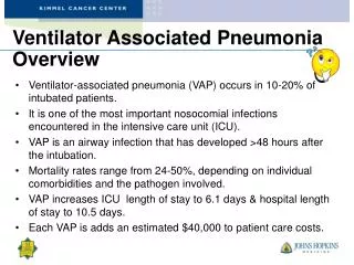
Why is it important • 10-20% of patients requiring mechanical ventilation will develop VAP. • Between 250,000 and 300,000 cases per year occur in the United States alone • Increased morbidity and mortality • VAP is associated with 15% of all nosocomial infections and 25% of the deaths associated with nosocomial infections. • Higher costs • 40,000 dollars to 57,000 dollars more than a patient that does not develop VAP (Koeing , Truwit, 2006)
What is VAP? • “VAP( Ventilator-Associated Pneumonia) is defined as a pneumonia occurring in patients requiring a device intermittently or continuously to assist respiration through a tracheostomy or endotracheal tube.” (“Safe Health Care”, 2007) • “Further, the device must have been in place within the 48 hour period before onset of infection and for at least 2 consecutive days.” (“Safe Health Care”, 2007)
Signs and Symptoms Most common • Fever (>38°C or >100.4°F) • Rales or bronchial breath sounds • Tachycardia • New onset of purulent sputum, or change in character of sputum, or increased respiratory secretions, or increased suctioning requirements • Worsening gas exchange (e.g., O2 desaturations increased oxygen requirements, or increased ventilator demand) (CDC, 2013)
Diagnosis Commonly used VAP criteria include • new or progressive pulmonary infiltrate on chest radiograph • fever (greater than 38.3ºC) • leukocytosis • purulent tracheobronchial secretions
Prevention is Key!!! • Head of Bed Elevation >30 • Prophylaxis to reduce DVT and PUD • Daily interruptions of sedation and daily assessment of readiness for extubation • Subglottic Suctioning • CHX Swab
Early, single chlorhexidine application • Randomized controlled clinical trial • Purpose: investigate the effect of a single application of chlorhexidine (CHX) by swab on the development of (VAP) • 55.6% of the control patients developed pneumonia compared to 33.3% of the intervention patients. • Early, single application of CHX was found to reduce VAP (Grap, M, Munro , Hamilton , Elswick, , Sessler, , & Ward, 2011)
Treatment • Prompt initiation of antibiotic therapy is a cornerstone of treatment of VAP • However, when VAP is first suspected, the bacteria causing infection is typically not known • Broad-spectrum antibiotics are given until the particular bacterium and its sensitivities are determined.
CASE STUDY Admitting Note Patient ID# 76-12-00 Diagnosis: Coronary Atherosclerosis 78-year old male (DOB: 12/03/28) who after evaluation by cardiovascular surgery service on 4/29, was diagnosed with coronary artery disease. Admitted on 5/18 for elective surgery after an extensive pre-hospital multi-system work up. He has lost 30 pounds in the last 3 months. Cefazolinordered on call to the operating room. (L) peripheral IV inserted PMH: Hyperlipidemia, renal insufficiency, myocardial infarction, obesity, pneumonia and urinary tract infection, bilateral cataracts, 10 years ago, unstable angina.
Case Study (Con) Admission Vital Signs & Labs: BP 130/70, P 88, R 20, Temp 37.1, Na 135, K 3.8, BUN 15, Cr 1.5, WBC 8.7, HCT 36 Surgical Procedure: Coronary Artery Bypass Graft using (L) was performed on 5/18 while the patient was under general anesthesia. Duration: 4 hours and 10 minutes. Admitted to CTICU on 5/18
5/18Afebrile, Lungs clear; intubated. (RIJ) internal jugular IV access device inserted. Foley catheter draining clear yellow urine. 5/19– Temp 36.5; Bilateral rhonchi; Thin yellow blood-tinged secretions. Chest x-ray shows slight congestion with infiltrate in RLL 5/20– Temp 38.6, Incision dressings clean and dry; Labored respirations (R=36), BP-96/50. Decreased O2 saturation, CXR-opacity in RLL. Bilateral rales on rhonchi. Suctioned for thick tan secretions. Sputum and blood cultures sent for C&S. Sputum culture- gram positive cocci
YES, fever, purulent sputum The Facts: • Had been on a vent. within the last 48 hours • Febrile (38.6) • New onset of purulent sputum • Respiratory Distress (Rate= 36) • 2 CXRs with RLL consolidation
Ineffective Airway Clearancerelated to inflammation, the accumulation of secretions Impaired Gas Exchangerelated to alveolar capillary membrane changes Hyperthermia related to inflammatory processes Imbalanced Nutrition Less than body requirements Outcomes: -Afebrile; Effective Airway clearance; Optimal gas exchange, adequate oxygenation to the tissue; Meet the needs of adequate nutrition
Prognosis Late-onset VAPhas poor prognosis in terms of mortality (66%) as compared to the early-onset type (20%) (Hina,Arun,Akhya, 2010) Case Study continued Started on IV antibiotics q6h on 5/21 Began weaning trials. Patient alert. Respiratory secretions decreased. Improved respiratory status. Extubated on 5/22 and transferred to 3 east next day.
NCLEX You are caring for a patient with emphysema and respiratory failure who is receiving mechanical ventilation through an endotraceal tube. To prevent ventilator-associated pneumonia (VAP), which action is most important to include in the plan of care? 1. Administer ordered antibiotics as scheduled2. Hyperoxygenate the patient before suctioning3. Maintain the head of the bed at a 30 - to 45-degree angle4. All of the Above
Conclusion • Prevention is Key! • Treatment: Prompt initiation of antibiotic therapy • S/S: Fever (>38°C or >100.4°F) , Ralesor bronchial breath sounds, Tachycardia, worsening gas exchange • Prognosis: Late-onset VAP has poor prognosis in terms of mortality (66%) as compared to the early-onset type (20%)
References Safer Healthcare Now; Campaign, How –to guide: Prevention Ventilation-Associated Pneumonia. May 2007 p1-40. Grap, M., Munro, C., Hamilton, V., Elswick, R., Sessler, C., & Ward, K. (2011). Early, single chlorhexidine application reduces ventilator-associated pneumonia in trauma patients. Heart & Lung: The Journal Of Critical Care, 40(5), e115-e122 Gadani H, Vyas A, Kar AK. A study of ventilator-associated pneumonia: Incidence, outcome, risk factors and measures to be taken for prevention. Indian J Anaesth2010;54:535-40 Centers for Disease Control and Prevention. Guidelines for preventing health-care-associated pneumonia, 2003: recommendations of CDC and the Healthcare Infection Control Practices Advisory Committee. MMWR 2004;53(No. RR-3). http://www.hanys.org/ihi_campaign/upload/VanAntwerpen%20Case_Studies.pdf