Download
1 / 1
10 likes | 141 Views
m-ICTE 2003. G. DE LUCA, C. EVANGELISTA AND G. ZITO , Department of Physics, University of Bari , Italy. Objective. We realized. TEACHING THE PHYSICS OF THE EYE BY USING JAVA APPLETS. A WEB HYPERTEXT containing: a description of the eye in its anatomic, physiological and optical aspects

E N D
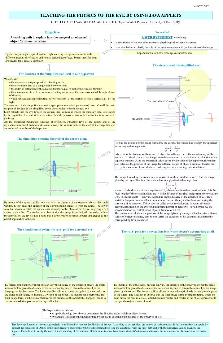
m-ICTE 2003 G. DE LUCA, C. EVANGELISTA AND G. ZITO, Department of Physics, University of Bari, Italy Objective We realized TEACHING THE PHYSICS OF THE EYE BY USING JAVA APPLETS • A WEB HYPERTEXTcontaining: • a description of the eye in its anatomic, physiological and optical aspects • java simulations to clarify the role of the eye’s components in the formation of the image A teaching path to explain how the image of an observed object forms on the retina http://www.ba.infn.it/%7eevangel/deluca/tesi.html Eye is a very complex optical system. Light entering the eye meets media with different indexes of refraction and several refracting surfaces. Some simplifications are needed for a didactic approach The structure of the simplified eye The features of the simplified eye used in our hypertext • We consider: • the cornea as a unique spherical refracting surface; • the crystalline lens as a unique thin biconvex lens; • the index of refraction of the aqueous humour equal to that of the vitreous humour; • the curvature centres of the various refracting surfaces on the same axis, called the optical axis of the eye; • valid the paraxial approximation, so we consider flat the portion of eye's surfaces hit by the light. • The structure of the simplified eye (with appropriate numerical parameters) “works” well, because the path of the light in the simplified eye is very similar to that in the real eye. • Light refracts into the eye through the cornea, then, coming in trough the pupillary hole, is refracted by the crystalline lens and strikes the retina; here the photosensitive cells transfer the information to the brain. • All the numerical parameters (indexes of refraction, curvature rays of the cornea and of the crystalline lens, focal distances, distances among the various parts of the eye) of the simplified eye are collected in a table of the hypertext The same index of refraction 3D vision The simulation showing the role of the cornea alone To find the position of the image formed by the cornea, the student has to apply the spherical refracting surface equation: where o is the distance of the observed object from the eye, r is the curvature ray of the cornea, i is the distance of the image from the cornea and n is the index of refraction of the aqueous humour. Using the numerical values given by the table of the hypertext, the student can calculate the position of the image for different values of object's distance, then he can verify the exactness of his calculus visualizing the corresponding Java simulation The image formed by the cornea acts as an object for the crystalline lens. To find the image given by the crystalline lens, the student has to apply the thin lens equation: where o is the distance of the image formed by the cornea from the crystalline lens, f is the focal length of the crystalline lens and i is the distance of the final image from the crystalline lens. The focal length f can vary depending on the distance of the observed object. This variation happens because ciliary muscles can contract the crystalline lens, so varying the curvature of its surfaces. This process is called accommodation and happens in various degrees, depending on the eye condition being normal or defective. For a normal eye the accommodation is greatest for an object's distance of 25 cm. The student can calculate the position of the image (given by the crystalline lens) for different values of object's distance, then he can verify the exactness of his calculus visualizing the corresponding Java simulation By means of the upper scrollbar one can vary the distance of the observed object: the small window below gives the distance of the corresponding image Ic from the retina. The lower scrollbar allows to rotate the optical axis normally to the plane of the figure, so giving a 3D vision of the effect. The student can observe that the image forms behind the retina, where the zone hit by the rays is not a point but a circle, which becomes greater and greater as the object approaches to the eye The simulation showing the rays’ path for a normal eye The rays’ path for a crystalline lens which doesn’t accomodate at all By means of the upper scrollbar one can vary the distance of the observed object: the small window below gives the distance of the corresponding image I from the retina. Ic is the image given by the cornea. The lower scrollbar allows to rotate the optical axis normally to the plane of the figure, so giving a 3D vision of the effect. The student can observe that the final image forms on the retina whatever is the distance of the object: this happens thanks to the accommodation process of the crystalline lens. By means of the upper scroll bar one can vary the distance of the observed object: the small window below gives the distance of the corresponding image I from the retina. Ic is the image given by the cornea. The lower scrollbar allows to rotate the optical axis normally to the plane of the figure. The student can observe that the final image forms behind the retina, where the zone hit by the rays is a circle, which becomes greater and greater as the object approaches to the eye: the object is seen blurred • The hypertext also contains: • an applet showing how the eye determines the direction under which an object is seen; • two applets illustrating the methods used by the eye to determine the distance of the observed object. The developed material reveals a great help to traditional lessons on the Physics of the eye. According to our opinion, the reason of such a success is this: the student can apply by himself the equations of Optics to the simplified eye and compare the results obtained (solving the equations) with the rays' path and with the numerical values given by the applets. This allows to verify the correct understanding of Geometrical Optics in a situation that attracts students’ attention and interest because concerns phenomena of everyday life.