Download
1 / 6
60 likes | 74 Views
Our team recently worked on a research paper, in a single-center case series, we report five cases of miscarriage after implantation of embryos with mosaicism of chromosome 20. The female partners were of advanced maternal age and had a history of previous miscarriages. The coupleu2019s blood karyotype reports were normal and there was no family history of any inherited disease.

E N D
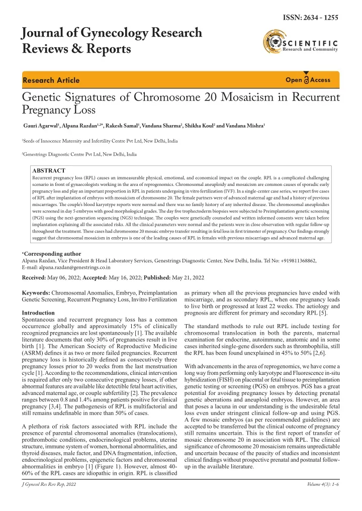
ISSN: 2634 - 1255 Journal of Gynecology Research Reviews & Reports Research Article Genetic Signatures of Chromosome 20 Mosaicism in Recurrent Pregnancy Loss Gauri Agarwal1, Alpana Razdan1,2*, Rakesh Samal1, Vandana Sharma2, Shikha Koul2 and Vandana Mishra1 Open Access 1Seeds of Innocence Maternity and Infertility Centre Pvt Ltd, New Delhi, India 2Genestrings Diagnostic Centre Pvt Ltd, New Delhi, India ABSTRACT Recurrent pregnancy loss (RPL) causes an immeasurable physical, emotional, and economical impact on the couple. RPL is a complicated challenging scenario in front of gynaecologists working in the area of reprogenomics. Chromosomal aneuploidy and mosaicism are common causes of sporadic early pregnancy loss and play an important proportion in RPL in patients undergoing in vitro fertilization (IVF). In a single-center case series, we report five cases of RPL after implantation of embryos with mosaicism of chromosome 20. The female partners were of advanced maternal age and had a history of previous miscarriages. The couple’s blood karyotype reports were normal and there was no family history of any inherited disease. The chromosomal aneuploidies were screened in day 5 embryos with good morphological grades. The day five trophectoderm biopsies were subjected to Preimplantation genetic screening (PGS) using the next-generation sequencing (NGS) technique. The couples were genetically counseled and written informed consents were taken before implantation explaining all the associated risks. All the clinical parameters were normal and the patients were in close observation with regular follow-up throughout the treatment. These cases had chromosome 20 mosaic embryo transfer resulting in fetal loss in first trimester of pregnancy. Our findings strongly suggest that chromosomal mosaicism in embryos is one of the leading causes of RPL in females with previous miscarriages and advanced maternal age. *Corresponding author Alpana Razdan, Vice President & Head Laboratory Services, Genestrings Diagnostic Center, New Delhi, India. Tel No: +919811368862, E-mail: alpana.razdan@genestrings.co.in Received: May 06, 2022; Accepted: May 16, 2022; Published: May 21, 2022 Keywords: Chromosomal Anomalies, Embryo, Preimplantation Genetic Screening, Recurrent Pregnancy Loss, Invitro Fertilization as primary when all the previous pregnancies have ended with miscarriage, and as secondary RPL, when one pregnancy leads to live birth or progressed at least 22 weeks. The aetiology and prognosis are different for primary and secondary RPL [5]. Introduction Spontaneous and recurrent pregnancy loss has a common occurrence globally and approximately 15% of clinically recognized pregnancies are lost spontaneously [1]. The available literature documents that only 30% of pregnancies result in live birth [1]. The American Society of Reproductive Medicine (ASRM) defines it as two or more failed pregnancies. Recurrent pregnancy loss is historically defined as consecutively three pregnancy losses prior to 20 weeks from the last menstruation cycle [1]. According to the recommendations, clinical intervention is required after only two consecutive pregnancy losses, if other abnormal features are available like detectible fetal heart activities, advanced maternal age, or couple subfertility [2]. The prevalence ranges between 0.8 and 1.4% among patients positive for clinical pregnancy [3,4]. The pathogenesis of RPL is multifactorial and still remains undefinable in more than 50% of cases. The standard methods to rule out RPL include testing for chromosomal translocation in both the parents, maternal examination for endocrine, autoimmune, anatomic and in some cases inherited single-gene disorders such as thrombophilia, still the RPL has been found unexplained in 45% to 50% [2,6]. With advancements in the area of reprogenomics, we have come a long way from performing only karyotype and Fluorescence in-situ hybridization (FISH) on placental or fetal tissue to preimplantation genetic testing or screening (PGS) on embryos. PGS has a great potential for avoiding pregnancy losses by detecting prenatal genetic aberrations and aneuploid embryos. However, an area that poses a lacuna in our understanding is the undesirable fetal loss even under stringent clinical follow-up and using PGS. A few mosaic embryos (as per recommended guidelines) are accepted to be transferred but the clinical outcome of pregnancy still remains uncertain. This is the first report of transfer of mosaic chromosome 20 in association with RPL. The clinical significance of chromosome 20 mosaicism remains unpredictable and uncertain because of the paucity of studies and inconsistent clinical findings without prospective prenatal and postnatal follow- up in the available literature. A plethora of risk factors associated with RPL include the presence of parental chromosomal anomalies (translocations), prothrombotic conditions, endocrinological problems, uterine structure, immune system of women, hormonal abnormalities, and thyroid diseases, male factor, and DNA fragmentation, infection, endocrinological problems, epigenetic factors and chromosomal abnormalities in embryo [1] (Figure 1). However, almost 40- 60% of the RPL cases are idiopathic in origin. RPL is classified J Gynecol Res Rev Rep, 2022 Volume 4(3): 1-6
Citation: Gauri Agarwal, Alpana Razdan, Rakesh Samal, Vandana Sharma, Shikha Koul (2022) Genetic Signatures of Chromosome 20 Mosaicism in Recurrent Pregnancy Loss. Journal of Gynecology Research Reviews & Reports. SRC/JGRRR-170. DOI: doi.org/10.47363/JGRRR/2022(4)151 conditions as per recommendations and after obtaining patient’s consent. During the course of pregnancy, ultrasonography was used to observe the status of Crown Rump Length (CRL), Nuchal Translucency (NT) and nasal bone. Maternal serum concentrations of free beta (β)-hCG biomarker were checked and regular follow- up was done. Informed consent was obtained from the individual(s) for the transfer of embryos and for publication of any potentially identifiable images or data in this study. Whole-Genome Amplification (WGA) Pre-Implantation Genetic Screening for Aneuploidy (PGS) was performed on day-5 biopsied embryos using the standard operating protocol of NGS. DNA was extracted from the trophectoderm, followed by whole genome amplification (WGA) using Ion Torrent Ion SingleSeqTM96 kit (Thermo Fisher Scientific, USA). The DNA libraries were purified and pooled followed by gel electrophoresis or DNA concentration was measured using QubitTM dsDNA HSkit, for quality control. Ion chef loading was done using the required chip (as required) after diluting the purified and pooled library. The NGS procedure was completed using the Ion Chef System (Thermo Fisher Scientific, USA) and Ion S5 sequencer (Thermo Fisher Scientific, USA). The data analysis was performed using Ion Reporter Software 5.10.5.0 using preconfigured workflow ReproSeq Mosaic PGS w1.1. Figure 1: Genetic and Nongenetic factors causing Recurrent Pregnancy Loss (RPL) and its Management Studies have reported chromosome 20 trisomy and monosomy in amniotic fluid and in chronic villi samples in association with no gross anomalies and few showing association with RPL [7-11]. Very few reports have found a special chromosome anomaly Ring chromosome 20, in association with the development of neurological disorders such as refractory epilepsy [12]. Chromosome 20 trisomy is a condition in an embryo (or an individual) with an extra chromosome 20 copy or a partial extra copy of chromosome 20 in a few or all cells. The chromosome 20 trisomy is one of the most common types of chromosomal abnormality that is frequently found during prenatal diagnosis. Studies have shown that the fetus was usually normal in prenatally diagnosed individuals, however, there are many phenotypically abnormal features in case of chromosome trisomy 20 such as epilepsies, skeletal abnormalities, heart defects, spinal abnormalities, hypotonia, chronic constipation, sloped shoulders, and learning deficits. The trisomy 20 (gain) error develops during the oocyte or sperm cells formation before fertilization, and mosaic trisomy 20 develops during cell division soon after fertilization [10,11]. Results High-resolution PGS by NGS has been used to reliably detect mosaic samples with 20-80% aneuploid cells. The female partners were aged between 32-42years, with weights ranging from 55- 68Kg, with North Indian ethnicity, poor ovarian reserve, no history of smoking, no diabetes, and no previous family history of inherited disease. The couple karyotypes were normal females having 46XX and males having 46XY. All patients had previous history of miscarriages. The patients underwent IVF treatment and day-5 embryo biopsies were subjected to PGS. The age of female partners and embryos screened for PGS with their clinical outcome have been detailed in Table 1 and a whole-genome view picture representing the mosaic gain on one of the chromosome 20 have been shown in figure 2. The pregnancy loss occurred in the first trimester of pregnancy. Since the clinical significance of chromosome 20 mosaicism embryo transfer remains uncertain. The present case series highlights chromosome 20 mosaicism associated with RPL in females with advanced maternal age and previous history of miscarriages. Case1 A 32-year female presented with infertility of 6years duration and previous pregnancy losses. The couple had normal karyotype reports and screen results were negative for antiphospholipid antibodies (APLA). Evaluation of all the clinical reports by the concerned gynecologist of the couple found moderate Oligoasthenoteratozoospermia (OATS) in the partner hence, the decision for ICSI was taken. The patient conceived with the transfer of a single embryo in the first cycle without PGS done on the embryo (as their decision). Unfortunately, this resulted in miscarriage in the 6th week. The decision for PGS was taken because 4 more potential embryos were remaining. Due to the acceptable transfer protocol of mosaic embryos as per recommendations of the Preimplantation Genetic Diagnosis International Society (PGDIS), the embryo with mosaic gain in chromosome 20 was selected and the decision for transfer was taken13. Though the patient conceived and was uneventful in the initial few weeks it resulted in a miscarriage at the 7th week of pregnancy. Materials and Methods Stimulation Protocol, Biopsy and Vitrification The couples were treated at our center (between the years 2020August-2022 March) with a previous history of recurrent miscarriages, and underwent IVF treatment with PGS. In all these cases controlled ovarian stimulation (COS) was done by short antagonist protocol. The dose of gonadotropin was calculated (based on anti-mullerian hormone) and was administered between 150-300U. Egg aspiration was done under total intravenous anaesthesia after 36 hours of Human chorionic gonadotropin (HCG trigger). Intracytoplasmic sperm injection (ICSI) was used and a fertilization check was done after ~18 hours. Zygotes were cultured in media (recommended) for the 5th day and blastocysts were graded according to institutional protocol. The resulting day- 5 embryos of good morphological grades (5AA,4AA,5AB,5BA) were biopsied where many trophectoderm cells (5-8 cells) by using a laser were taken, collected in sterile phosphate buffer saline (1X) (Affymetrix) and stored at -200C. The vitrification method was used to freeze the embryos. These were further subjected to PGS based on NGS assay. The trophectoderm DNA was then screened for genetic abnormalities, and only the compatible embryos were transferred into the uterine cavity under strict quality control Case 2 Another couple profile with a history of previous 3 abortions at approximately 6-8 weeks with normal karyotype and negative Antiphospholipid antibodies (APLA) screen underwent IVF. The OCS was performed and taken for ICSI. The trophectoderm J Gynecol Res Rev Rep, 2022 Volume 4(3): 2-6
Citation: Gauri Agarwal, Alpana Razdan, Rakesh Samal, Vandana Sharma, Shikha Koul (2022) Genetic Signatures of Chromosome 20 Mosaicism in Recurrent Pregnancy Loss. Journal of Gynecology Research Reviews & Reports. SRC/JGRRR-170. DOI: doi.org/10.47363/JGRRR/2022(4)151 biopsied embryos were subjected to PGS. The option was to transfer the embryo with chromosome 20 mosaicism. Initially, there was no complication, and the patient conceived which resulted in a miscarriage in the 8th week. Table 1: Patient details and Clinical outcome of Pregnancy Case Number Patient’s Age (Years) Oocyte source Total Number of embryos screened for PGS Mosaicism (% gain) Miscarriage at week P-1 32 Self 5 Chr 20 (45%) 7th Case 3 The couple was presented with previous 2 miscarriages at 12 weeks and 8 weeks respectively for unexplained reasons and came to our center for IVF treatment. Cytogenetic analysis of couple of karyotypes were normal and there was no family history of inherited disease. IVF treatment was initiated, OCS was performed and ICSI was used. The trophectoderm biopsied embryos were subjected to PGS and results showed most of the embryos were aneuploid and one with chromosome 20 mosaicism. Therefore, the decision was taken and the patient was counseled detailing all the consequences of embryo transfer with mosaicism and the risk of miscarriage. The transfer was made and the patient conceived but was unsuccessful and the fetus could not survive beyond the 6th week even after close observation. P-2 35 Self 8 Chr 20 (25%) 8th P-3 36 Self 6 Chr 20 (35%) 6th P-4 37 Self 6 Chr20 (25%) 5.5 P-5 42 Self 3 Chr 20 (35%) 7.2 Discussion Several factors of genetic, autoimmune, and environmental play an important role in the pathophysiology of RPL (Figure-1). Maternal age is one of the crucial factors, related to the increase in the frequency of abnormal or mosaic embryos [2]. The quality of the oocyte, embryo, its morphological grade, and many other parental factors may cause RPL. The errors in meiosis resulting in aneuploidy in embryos are the possible reasons behind failed implantation, miscarriages, and congenital birth defects as reported previously [14,15]. The available literature documented that postzygotic, mitotic nondisjunction events result in presence of both normal, abnormal, or mosaic cell lines. Although there is no such study prospective or retrospective in the literature, we report it for the first time. The mosaicism of chromosome 20 results in significant changes in clinical and phenotypic expression varying from individual to individual depending on the degree of mosaicism. Figure 2: Whole-Genome view showing Chromosome 20 Mosaicism (gain) in one of the cases Case 4 The female patient was of 37-year-old with a history of IVF failure with 2 unexplained miscarriages previously. The couple’s karyotype was normal and there was no family history of an inherited disease. The couple came to our fertility center seeking fertility treatment. The female partner had no known allergies, no sexually transmitted diseases, and was negative for APA. All routine tests and clinical parameters were checked and IVF treatment was initiated. The day-5 embryo biopsy was subjected to PGS. The embryo with chromosome 20 mosaicism was transferred after taking informed consent and a counseling session completed with the couple. Initially, the pregnancy was uneventful but the fetus could not survive beyond the 5.5 weeks. There are a few studies explaining abnormal chromosome 20 (aneuploid, trisomy 20, monosomy 20) in association with developmental delays, miscarriages, and epilepsy respectively [16,17]. Mosaicism with less than 40% levels of trisomy 20 in amniotic fluid (<40% trisomic cells) were associated with a lower risk for abnormalities [18]. An association between the level of trisomy and outcome, with only 4% abnormal outcomes when <40% trisomic cells were reported [18]. However, clinical trial or case reports including transfer of embryo with varying degrees of mosaicism of chromosome 20 in association with prenatal or postnatal follow-up are lacking. In the present study, we report five cases in which chromosome 20 (mosaic) embryos were implanted, resulting in unsuccessful pregnancy and miscarriages within 6 to 8 weeks. The PGS was performed to screen the chromosomal aneuploidies in cells before the implantation of embryos. The clinical indications common in our patients were advanced maternal age (except one patient), poor ovarian reserve, recurrent miscarriages, recurrent implantation failures, and previous abnormal pregnancy. Cytogenetic reports of couple karyotyping were normal in all the cases and there was no family history of an inherited disorder. Case 5 The mother aged 42 years had previous IVF failures, had Polycystic ovary syndrome (PCOS), and had previous miscarriages. The couple had normal karyotype reports with no family history of any inherited disease. The IVF treatment was initiated. The embryo biopsies were subjected to PGS to screen for chromosomal aneuploidies. None of the embryos were euploid, the only choice was one embryo with mosaicism of chromosome 20 and transferred after counseling and obtaining informed consent from the patient. Post transfer the pregnancy was without any complications however, the fetal loss was reported at the 7.2th week of pregnancy in this case. We observed embryos implanted with chromosome 20 mosaicism (gain) resulted in fetal loss. The fetal demise was diagnosed by ultrasound scan and the gestational age at the time of miscarriage was estimated in all the patients under close observation and follow- up. In our study, one patient was having PCOS, with previous miscarriages. PCOS has been reported to be associated with an increased risk of miscarriages, in approximately 25% of cases either following spontaneous conception or ovulation induction J Gynecol Res Rev Rep, 2022 Volume 4(3): 3-6
Citation: Gauri Agarwal, Alpana Razdan, Rakesh Samal, Vandana Sharma, Shikha Koul (2022) Genetic Signatures of Chromosome 20 Mosaicism in Recurrent Pregnancy Loss. Journal of Gynecology Research Reviews & Reports. SRC/JGRRR-170. DOI: doi.org/10.47363/JGRRR/2022(4)151 previously [19,20]. The possible explanation for RPL in these cases is advanced maternal age, previous history of miscarriage, and PCOS in one case. Epidemiological studies have related advanced maternal age as one of the strong risk factors for the increase in chromosomal mosaicism and abnormalities [21, 22]. The authors claimed that the frequency and distribution of cytogenetically abnormal miscarriages in couples with spontaneous abortions or recurrent miscarriages were more among females with advanced age [21]. The rate of aneuploid embryos and miscarriages were significantly higher among these with a maximum gestational age at the time of fetal loss <2 weeks [21]. In a metanalysis, an association between an abnormal outcome in 50% (5/10) of the pregnancies with higher than 80% trisomy 20 cells, 28% (17/61) of the pregnancies with greater than 40% trisomy 20 cells, and 4% (8/201) of the pregnancies with less than 40% trisomy 20 cells, has been documented [18]. However, provided a contradictory approach and stated that trisomy mosaicism levels do not influence the outcome in prenatally detected mosaic trisomy 20, and therefore, a second amniocentesis is needed along with genetic counselling [31,32]. It has been hypothesized that the maximum chromosomal abnormalities observed/ found in spontaneous miscarriages occur de novo and are due to random errors that occurred at the time of gametogenesis and embryonic development [33]. The risk of a fetal trisomy increases with advanced maternal age [34,35]. Since the average age of women bearing their first child has increased over the last two decades in developing as well as developed countries, chromosomal anomalies have likely to become more frequent and prevalent [34]. It has been well established previously that the mosaic embryo transfer is an alternative option only for patients in absence of any euploid embryo indicated from PGS, and should be opted only after comprehensive genetic counseling, patient’s consent, and follow-up along with recommended prenatal diagnosis [23]. Previous miscarriages have also been one of the major risk factors which might have increased the susceptibility to miscarriage. In our study, all the cases had a history of miscarriages. It has been reported that the risk of further fetal loss increases to approximately 50% among females with three or more miscarriages without a liveborn infant [24-26]. Mis-segregation errors at the time of fertilization and impaired cleavage morphokinetics can result in an embryo with chromosomal abnormalities. Studies focussed on mosaicism of chromosome 20 trisomy have shown that the infant born were normal in the vast majority of prenatally diagnosed cases [36]. However, abnormalities reported included spinal problems (including spinal stenosis, vertebral fusion, and kyphosis), hypotonia (decreased muscle tone), lifelong constipation, sloped shoulders, and significant learning disabilities despite normal intelligence [36]. Although the risk for congenital abnormalities in a low level of trisomic cells is less than 10%, parents should be advised of additional risks, such as neurodevelopmental delay and neuropsychiatric manifestations [37]. Since the molecular mechanism underlying RPL remains uncertain we hypothesize that the increased stress on the endometrium of the uterus and de novo genetic variants and nondisjunction events may be the causes for RPL in our cases. Although the endometrium has a good potential for plasticity and regeneration, the repeated events hamper the endometrium to prepare for pregnancy or decidualization [27]. Probably the persistence of miscarriages even after any conception cycles might have resulted in successive pregnancy loss. Additionally, the mosaic embryos might have not met the mechanism of self-correctness and did not survive. Conclusion RPL continues to be a challenge for patients as well as for gynaecologists. Therefore, ruling out the underlying reasons including embryo abnormalities can be of significance in decision making and management of RPL. Differences in the level of mosaicism of chromosome 20 may cause a remarkable variation in the clinical symptoms and phenotypic expression varying from individual to individual. In our case series, mosaicism was associated with poor clinical outcomes and RPL. The possibility of pregnancy post transfer of chromosome 20 (mosaicism), its capability to self-correct and the associated uncertain clinical outcome must be considered carefully along with the clinical recommendations. The decision to utilize embryos that are not clearly euploid (or mosaic or aneuploid) must be discussed thoroughly among the diagnostic team, genetic counsellor, and concerned gynaecologist and patient or couple undergoing IVF treatment. Our study confirms that chromosomal abnormality especially the chromosome 20 mosaicism is one of the reasons contributing to RPL in patients with advanced maternal age and with a history of RPL. Chromosomal analysis or whole-genome analysis is a must to be done part to rule out the aetiology among couples with recurrent miscarriages in order to avoid the unfavorable outcome. The prenatally diagnosed mosaicism of chromosome 20 needs to be evaluated and investigated further in prospective cohort studies in order to alter the odds of RPL. The transfer recommendations should be followed along with comprehensive parental counseling advising on the risk associated such as developmental delay and miscarriages. We strongly recommend considering all the important clinical, genetic and nongenetic factors along with guidelines while deciding on the transfer of mosaic embryos to optimize and manage the favorable outcome in cases with RPL. It has been observed previously that in couples who had repeated miscarriages or recurrent implantation failure after IVF, the proportion of chromosomal disturbances was found to be accentuated [28]. Our findings are in concordance with previous reports. A study reported seven spontaneous abortions with non-mosaic trisomy 20 associated with miscarriages and evaluated meiotic origin in five cases (three maternal and two undetermined) and probable somatic origin in two cases [29]. An extra copy of chromosome 20 in all the cells is a rare condition, and a fetus with this chromosomal abnormality does not survive past the first trimester of pregnancy [16]. Trisomy 20 mosaicism is found in prenatal diagnosis, and the rate of a poor outcome was 6.5% with wide variability in phenotype [7]. The liveborn were observed with abnormalities such as growth retardation, hypotonia, structural and neurological abnormalities and seizures, facial dysmorphism, failure to thrive, and developmental delay. The majority of reported studies have found that miscarriages occur in chromosomally normal parents due to abnormal chromosomes in the implanted embryo. However, balanced chromosome abnormalities have been reported among 2–5% of patients with recurrent miscarriages [30]. In our study, all the patients were normal as clear from available peripheral blood karyotype reports. J Gynecol Res Rev Rep, 2022 Volume 4(3): 4-6
Citation: Gauri Agarwal, Alpana Razdan, Rakesh Samal, Vandana Sharma, Shikha Koul (2022) Genetic Signatures of Chromosome 20 Mosaicism in Recurrent Pregnancy Loss. Journal of Gynecology Research Reviews & Reports. SRC/JGRRR-170. DOI: doi.org/10.47363/JGRRR/2022(4)151 Conflicts of Interest The authors declare no conflicts of interest. et al. (2005) Prenatally detected trisomy 20 mosaicism. Prenat Diagn 25: 239-4. 19. Revised 2003 consensus on diagnostic criteria and long-term health risks related to polycystic ovary syndrome (2004) Rotterdam ESHRE/ASRM-Sponsored PCOS Consensus Workshop Group. Fertil Steril 1:19-25. 20. Glueck CJ, Wang P, Bornovali S, Goldenberg N, Sieve L (2003) Polycystic ovary syndrome, the G1691A factor V Leiden mutation, and plasminogen activator inhibitor activity: association with recurrent pregnancy loss. Metabolism 52: 1627-1632. 21. Choi HW, Yong-Seog P, Sun-Hee L, Lim CK, Seo JT, et al. (2016) Effects of maternal age on embryo quality and pregnancy outcomes using testicular sperm with intracytoplasmic sperm injection. Clinical and Experimental Reproductive Medicine 43: 221-227. 22. Sullivan AE, Silver RM, Lacoursiere DY, Porter TF, Branch DW (2004) Recurrent fetal aneuploidy and recurrent miscarriage. Obstet Gynecol 104: 784-788. 23. Grati FR (2014) Chromosomal Mosaicism in Human Feto- Placental Development: Implications for Prenatal Diagnosis. J Clin Med 3: 809-837. 24. Poland BJ, Miller JR, Jones DC, Trimble BK (1977) Reproductive counseling in patients who have had a spontaneous abortion. Am J Obstet Gynecol 127: 685-691. 25. Regan L, Braude PR, Trembath PL (1989) Influence of past reproductive performance on risk of spontaneous abortion. BMJ 299: 541-545. 26. Rubio C, Pehlivan T, Rodrigo L, Simon C, Remohi J, et al. (2005) Embryo aneuploidy screening for unexplained recurrent miscarriage: a minireview. Am J Reprod Immunol 53:159-165. 27. Lucas ES, Dyer NP, Murakami K, Lee YH, Yi-Wah C, et al. (2016) Loss of Endometrial Plasticity in Recurrent Pregnancy Loss. Stem Cells 34: 346-356. 28. WHO (1977) Recommended definitions, terminology and format for statistical tables related to the perinatal period and use of a new certificate for cause of perinatal deaths. Modifications recommended by FIGO as amended October 14, 1976. Acta Obstet Gynecol Scand 56: 247-253. 29. Robinson WP, Bernasconi F, Lau A, McFadden DE (1999) Frequency of meiotic trisomy depends on involved chromosome and mode of ascertainment. Am J Med Genet 84: 34-42. 30. Priya PK, Mishra VV, Roy P, Patel H (2018) A Study on Balanced Chromosomal Translocations in Couples with Recurrent Pregnancy Loss. J Hum Reprod Sci 11:337-342. 31. Bianca S, Ingegnosi C, Tetto C, Cataliotti A, Ettore G (2005) Prenatally detected trisomy 20 mosaicism and genetic counseling. Prenat Diagn 25: 725-726. 32. Bianca S, Boemi G, Barrano B, Cataliotti A, Ingegnosi C, et al. (2008) Mosaic trisomy 20: considerations for genetic counseling. Am J Med Genet 146: 1897-1898. 33. Carvalho B, Dória S, Ramalho C, Brandão O, Sousa M, et al. (2010) Aneuploidies detection in miscarriages and fetal deaths using multiplex ligation-dependent probe amplification: an alternative for speeding up results?. Eur J Obstet Gynecol Reprod Biol 153: 151-155. 34. Hassold, D (1985) Chiu Maternal age-specific rates of numerical chromosome abnormalities with special reference to trisomy. Hum. Genet 70: 1-17. 35. Franasiak JM, Forman EJ, Hong KH, Werner MD, Upham KM, et al. (2014) The nature of aneuploidy with increasing age of the female partner: a review of 15,169 consecutive References 1. Ford HB, Schust DJ (2009) Recurrent Pregnancy Loss: Etiology Diagnosis, and Therapy. Rev Obstet Gynecol Spring 2: 76-83. 2. Practice Committee of the American Society for Reproductive Medicine (2012) Evaluation and treatment of recurrent pregnancy loss a committee opinion. Fertil Steril 98: 1103- 1111. 3. Bender Atik R, Christiansen OB, Elson J, Kolte AM, Lewis S, et al. (2018) ESHRE guideline: Recurrent pregnancy loss. Human Reproduction Open 2: hoy004. 4. https://www.eshre.eu/Guidelines-and-Legal/Guidelines/ Recurrent-pregnancy-loss. 5. Egerup P, KolteAM, Larsen EC, Krog M, Nielsen HS, et al. (2016) Recurrent pregnancy loss: what is the impact of consecutive versus non-consecutive losses?. Hum Reprod 31: 2428-2434. 6. Hyde KJ, Schust DJ (2015) Genetic considerations in recurrent pregnancy loss. Cold Spring Harb Perspect Med 5: a023119. 7. Velissario V, Antoniadi T , Gyftodimou J , Bakou K , Grigoriadou M , et al. (2002) Maternal uniparental isodisomy 20 in a foetus with trisomy 20 mosaicism: clinical, cytogenetic and molecular analysis. European Journal of Human Genetics 10: 694-698. 8. Wallerstein R, Yu MT, Neu RL, Benn P, Lee Bowen C, et al. (2000) Common trisomy mosaicism diagnosed in amniocytes involving chromosomes 13, 18, 20 and 21: karyotype- phenotype correlations. Prenat Diagn 20: 103-122. 9. Hsu LYF, Kaffe S, Perlis TE (1991) A revisit of trisomy 20 mosaicism in prenatal diagnosis – an overview of 103 cases Prenat Diagn 11: 7-15. 10. Hayashi S, Miharu N, He H, Honda H, Samura O, et al. (1997) A phenotypically normal liveborn male after prenatal diagnosis of trisomy 20 mosaicism. J Obstet Gynaecol Res 23: 301-305. 11. Chen CP, Chang SD, Chueh HY, Su YN, Chern SR, et al. (2013) Discrepancy in the trisomy mosaicism level between cultured amniocytes and uncultured amniocytes in prenatally detected mosaic trisomy 20. Taiwan J Obstet Gynecol 52: 145-146. 12. Patil AA, Vinayan KP, Roy AG (2020) Epilepsy in Ring Chromosome 20 Syndrome Might have Variable Clinical Features. Ann Indian Acad Neurol 23: 718-722. 13. Gleicher N, Albertini DF, Barad DH, Homer H, Modi HD, et al. (2020) The 2019 PGDIS position statement on transfer of mosaic embryos within a context of new information on PGT-A. Reproductive Biology and Endocrinology 18: 57. 14. Martinez MC, Mendez C, Ferro J, Nicolas M, Serra V, et al. (2010) Cytogenetic analysis of early nonviable pregnancies after assisted reproduction treatment. Fertil Steril 93: 289-292. 15. Viotti M (2020) Preimplantation genetic testing for chromosomal abnormalities: aneuploidy, mosaicism, and structural rearrangements. Genes (Basel) 11: 602. 16. Mavromatidis G, Dinas K, Delkos D, Vosnakis C, Mamopoulos A, et al. (2010) Case of prenatally diagnosed non-mosaic trisomy 20 with minor abnormalities. Journal of Obstetrics and Gynaecology Research. August 36: 866-868. 17. Joo JG, Beke A, Toth-Pal E, Hargitai B, Szigeti Z, et al. (2006) Trisomy 20 mosaicism and nonmosaic trisomy 20: A report of 2 cases. J Reprod Med 51: 209-212. 18. Robinson WP, McGillivray B, Lewis ME, L Arbour, I Barrett, J Gynecol Res Rev Rep, 2022 Volume 4(3): 5-6
Citation: Gauri Agarwal, Alpana Razdan, Rakesh Samal, Vandana Sharma, Shikha Koul (2022) Genetic Signatures of Chromosome 20 Mosaicism in Recurrent Pregnancy Loss. Journal of Gynecology Research Reviews & Reports. SRC/JGRRR-170. DOI: doi.org/10.47363/JGRRR/2022(4)151 trophectoderm biopsies evaluated with comprehensive chromosomal screening. Fertil Steril 101: 656-663. 36. https://rarediseases.info.nih.gov/diseases/5332/chromosome- 20-trisomy/cases/28861. 37. Colizzi M, Antolini G, Passarella , Rizzo V, Puttini, et al. (2021) Additional Evidence for Neuropsychiatric Manifestations in Mosaic Trisomy 20: A Case Report and Brief Review. Children (Basel) 8: 1030. Copyright: ©2022 Alpana Razdan, et al. This is an open-access article distributed under the terms of the Creative Commons Attribution License, which permits unrestricted use, distribution, and reproduction in any medium, provided the original author and source are credited. J Gynecol Res Rev Rep, 2022 Volume 4(3): 6-6