Download
1 / 70
700 likes | 831 Views
Neurological Assessment and Disorders. Fetal Brain Development. The brain is one of the earliest organs formed during embryonic development Week 3 - beginning development of the brain, spinal cord, and heart

E N D
Fetal Brain Development • The brain is one of the earliest organs formed during embryonic development • Week 3 - beginning development of the brain, spinal cord, and heart • Weeks 4 to 5 - formation of tissue that develops into the vertebra and some other bones • At 7weeks the medulla, pons, and midbrain is formed • By 9 wks, the fetus will display spontaneous mvmt • 10 wks – begins to breathe • 13 wks - sucking motions are made with the mouth • 20 wks – responses to sound
Nutrition • Certain nutrients have a important roles in brain development; some have greater impact than others • Protein, fats, iron, zinc, iodine, vitamins A, B6 &K, folate • Any nutrient deficit effect is dependent on timing, dose and duration • Timing in terms of brain development process • Timing in terms of prevalence of nutrient deficit in population • Little evidence currently for enhancement of brain development in typically developing humans
Anatomy and Physiology of the Brain • Brain anatomy • Composed of 2 major areas • Cerebellum • Cerebrum
Anatomy and Physiology of the Brain • Cerebellum • Promotes intregrative muscle function • Maintains balance • Enables smooth, purposeful movements
Anatomy and Physiology of the Brain • Cerebrum • Central hemispheres with 4 lobes • Parietial • Frontal • Occipital • Temporal • Corpus Callosum – fiber bundles connecting the cerebral hemispheres • Cerebral Cortex • The ‘mind’, intellect • Grey matter • Third ventricle • Thalamus (integrates sensory input) • Hypothalamas (regulates body tempature)
Anatomy and Physiology of the Neurological System • Brain Anatomy • Brain stem • Relays input and output signals between the higher brain and the spinal cord. • It has three main components • The medulla, which controls areas of the abdomen, thorax, neck, throat, and mouth, and is part of cranial nerves VIII, IX, X, XI, and XII • The pons, which carries information between the brainstem and cerebellum • The midbrain, which is involved in eye movements
Glucose • Glucose metabolism • The neonatal brain is glucose dependent and the serum glucose provides the brain with a glucose pool. • The body preferentially pumps glucose against the gradient to the brain and cerebral metabolism is influenced by the availability of glucose and oxygen. • Anything that causes inadequate cerebral perfusion will compromise the glucose and oxygen supply to the brain. • Anaerobic metabolism causes lactic acid build-up and produces significantly smaller amounts of energy. • The premature infant has minimal-to-no glucogen stores and a less efficient glucose uptake mechanism.
Physiology of the Neurological System • Cerebral blood flow • Affected by pH, K, hypoxemia, osmolarity,and Ca • The brain will increase blood flow to spare itself inadequate supply of oxygen and electrolytes. • Low pH, hypoxia, hyperkalemia, and increased osmolarity will cause an increase in cerebral blood flow. • Increased Ca ion causes a decrease in cerebral blood flow.
Physiology of the Neurological System • Autoregulation • Maintains a steady-state of cerebral blood flow despite systemic blood pressure changes. • Limited or impaired autoregulation exists in premature or sick newborns. • Without autoregulation systemic blood pressure regulates cerebral perfusion. • Hypotension will lead to ischemia, which damages blood vessels, and surrounding tissues. • When adequate blood supply resumes, hemorrhage can occur into the ischemic areas. • Hypertension without autoregulation will increase cerebral flow rupturing blood vessels leading to hemorrhage
Determination of state, posture, and evaluation of movements Neurological Assessment
Assessment is based on gestational age Reflexes Root Suck (begins at ~32wks, intact by ~36wks) Moro Tonic neck Grasp (very strong in premmies) Babinski Hand- Mouth Others The presence or absence of these reflexes are indicative of: Gestational age Neurological abnormalities Progression of care Reflexes
Neurological Assessment • State • Deep sleep, drowsy, quiet-alert, active, crying, or active with stimulation • Posture • Posture is determined by gestational age • The more premature infant has extended, open posture, reflecting diminished tone. • The more term infant has flexed posture, reflecting adequate tone • Abnormal posture includes hyperextension, asymmetry, and flaccidity.
Neurological Assessment • Evaluation of movements • Should be symmetrical with smooth, coordinated quality. Note if jittery or tremulous, or if seizure activity is present. • Respiratory activity is observed for any signs of distress or apnea. • Cranial nerve function assessment. • Corneal reflex indicates intact cranial nerves V and VII. An absent corneal reflex with tonic neck reflex present is associated with severe brain damage • Blink reflex requires intact cranial nerves III and VII. Tongue movement, suck, swallow, gag, and cry are regulated by cranial nerves IX, X, XII • Muscle tone: Evaluate head lag, ventral suspension, clonus and recoil from extension. Check symmetry and briskness vs. flaccidity
Seizures • Result from excessive simultaneous electrical discharge or depolarization of neurons; can have many different causes. • Metabolic: Due to ischemia, hypoxemia, hypoglycemia, hypo- or hypernatremia, hypocalcemia, and hypomagnesemia. • Structural: Due to IVH, intrapartum trauma or hypoxic ischemic encephalopathy. • Intracerebral meningitis. • Withdrawal from maternal drug use
Incidence • Incidence of neonatal seizures in term vs preterm infants • 1.5-3.0 per 1000 live term births • 50-150 per 1000 live preterm births • Incidence as a function of birth weight • 57.5 per l000 in infants < 1500 grams • 2.8 per 1000 in infants 2500 to 3999 grams
Incidence • Precise frequency is difficult to delineate • Most of neonatal seizures we know is based on clinical observation • EEG monitoring with video recorder or direct inspection showed • Some clinical seizures do not have EEG correlation • Some electrographic seizures are not accompanied by clinical seizures
History • Family history may suggest genetic syndrome • Many of these syndromes are benign • In the absence of other etiologies, family history of seizures may suggest good prognosis • Pregnancy history is important • Search for history that supports TORCH infections, history of fetal distress, preeclampsia or maternal infections • Apgar scores offer some guidance • Low Apgar score without the need for resuscitation and subsequent neonatal intensive care is unlikely to be associated with neonatal seizures
Clinical Presentation May be subtle tonic, multifocal, focal clonic, or myoclonic seizure activity
Subtle Seizures • Selected Major Manifestations • Ocular phenomena • Tonic horizontal deviation of eyes with or without jerking of eyes • Sustained eye opening with ocular fixation • Oral-buccal-lingual movements • Chewing, sucking, lip smacking • Limb movements • Cycling, swimming, rowing • Autonomic phenomena • Increase in blood pressure, brady/tachycardia • Apneic spells
Clonic Seizures • Usually involve one limb or one side of the body jerking rhythmically at 1-4 times per second • Focal Clonic Seizures • Well localized clonic jerking • Infant usually not unconscious • Multifocal Clonic Seizures • Involves several parts of the body in migrating fashion (non-ordered fashion) • Simultaneous or in sequence
Focal and Multifocal Myoclonic • Focal and Multifocal Myoclonic Seizures • Well localized, single, multiple, migrating jerks usually of limbs • Usually not accompanied by EEG seizure discharges • Rare in neonates • Focal • Typically involve flexor muscle of upper extremity • Uncommon • Multifocal • Asynchronous twitching of several parts of the body • Characteristic in full term infants with hypoxic ischemic encephalopathy (HIE).
Tonic Seizures • Characteristic of infants < 2500 gms and seen with severe IVH • Focal Tonic Seizures • Sustained posturing of limb • Asymmetrical posturing of trunk and neck • Generalized Tonic Seizures • Tonic extension of upper and lower limbs (mimics decerebrate posturing) • Tonic flexion of upper limbs and extension of lower limbs (mimics decorticate posturing) • Most are not accompanied by EEG seizure discharges (only 30% have EEG correlates)
Seizures • Patient Care: • Positively identify movement as seizure activity by trying to stop movement with gentle passive flexion of the affected extremity • If the movement stops, it was not seizure activity • Check lab work; electrolytes, glucose, ABG, and septic work up. • Administer Phenobarbital or Valium as ordered. • Monitor oxygenation and vital signs. During a seizure, an infant’s blood pressure will rise, O2 saturation will drop, and CO2 will increase • Document carefully. • Educate and support the family.
Jitteriness vs Seizure Clinical Feature Jitteriness Seizure Abnormality of gaze or -- + eye movement Movements stimulus + -- sensitive Predominant movement Tremor Clonic jerking Movements cease with passive flexion + -- Autonomic changes -- + Volpe JJ. Neonatal seizures. In: Neurology of the Newborn 1995
Treatment • Ensure adequate ventilation and perfusion • If with hypoglycemia • Glucose 10% 2 ml/kg IV • If without hypoglycemia • Anticonvulsant therapy (Phenobarbital) • Etiology-Specific (as indicated) • Hypocalcemia (calcium gluconate 5% 4 ml/kg IV) • Hypomagnesemia (MgSO4 50% 0.2 ml/kg IV) • Pyridoxine deficiency (pyridoxine 50-100 mg IV) • Meningitis (Ampicillin/Cefotaxime
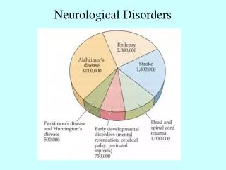
Neurological Disorders • Microencephaly • OFC is < 10% for gestational age • It is caused by a neuronal proliferation defect and occurs between 2 and 4 months gestation • The most severe cases occur in earlier gestation • Outcome is dependent on severity and may be associated with developmental delays
Neurological Disorders • Anencephaly - absence of neural tube closure exposing neural tissue • Occurs as a malfunction in the first state of neurological development,dorsal induction. • Infants lack brain above the brainstem and partial absence of skull bones with absent cerebrum. • May or may not lack cerebellum, brainstem and spinal cord. • Infants do not usually survive beyond the neonatal period. • Care includes comfort measures for the infant, and family support. • Encourage the family to see the infant and obtain genetic counseling for the family.
Neurological Disorders • Hydrocephaly • Congenital hydrocephalus is excess cerebral spinal fluid (CSF) in the ventricles of the brain. • CSF is produced from the parenchyma, cerebral ventricles, areas along the spinal cord, and the choroids plexus • Excessive CSF production, inadequate CSF absorption, or obstruction in the outflow tract can all result in hydrocephalus • Infants have a large head, separated sutures, full and tense fontanelles, increasing OFC, sunset eyes (signifies brain tissue damage) and visible scalp veins
Patient care Neurosurgery and genetic consults Placement of VP shunt, subgaleal shunt or reservoir Careful positioning of the head using cushions to prevent skin break down Normalize care as much as possible Review shunt placement and care with parents Post shunt placement Watch for signs of infection or blocked shunt, such as irritability, vomiting, increased head circumference, lethargy. Monitor for a change in feeding pattern. Assess for full or bulging fontanelles. Keep infant’s head off of the shunt site Neurological Disorders - Hydroencephaly
Neurological Disorders • Post-hemorrhagic hydrocephalus • The progressive dilatation of the ventricles following intraventricular hemorrhage (IVH). • Caused by periventricular white matter injury in the area of the germinal matrix. • Can be acute or chronic. • Acute rapidly appears within days of the initial bleed occurring secondary to malabsorption of CSF due to a clot in the ventricles. • Chronic is from inhibition of CSF flow or from blood from an ICH or pus from infection. The infants have a rapid increase in OFC, apnea, lethargy, increased intracranial pressure, and tense fontanelle, or may be asymptomatic. As progression occurs the signs of an acute bleed may become evident.
Neurological Disorders – Post Hemorrhagic Hydrocephalus • Patient care: • Daily OFC. • Serial head ultrasound. • Neurosurgery consult • Serial LP if indicated to decrease ICP or medications to decrease CSF production, such as Lasix or Diamox. Infant may need a ventroperitoneal shunt placed if hydrocephalus does not resolve. • Observe for signs of increased intracranial pressure and hydrocephalus
Neurological Disorders • Myelomeningocele • A neural tube defect that is a protrusion of the meninges lying directly under the skin, the internal surface of the spinal cord and or the nerve roots • It results from an error in dorsal induction • The majority of cases occur in the thoracolumbar, lumbar and lumbosacral regions. • There may be a herniated sac protruding from the back; it can be sealed or leaking • It is usually associated with hydrocephalus or Arnold-Chiari malformation, which involves some common features like reflux and aspiration, laryngeal stridor, central hypoventilation and apnea
Myelomeningocele • Pt Care • Examine and measure defect, note location and appearance. • Culture lesion of sac if open. • Wrap the lesion with sterile gauze moistened with warm NS. • Keep the infant in a prone position; place drape over the buttocks below the lesion to avoid contamination. • Consult a neurosurgeon for surgical closure of defect and a urologist to rule out neurogenic bladder • Follow developed protocol
Myelomeningocele • Most infants will have no significant mental retardation with varying degrees of paralysis of lower extremities. • Lesions below S-1: Children will learn to walk unaided. • Lesions between L-4 and L-5: Children will be able to walk with crutches. • Lesions above L-2: Children are usually wheel chair dependent.
Encephalocele • A neuronal herniation that may or may not contain meninges or brain parenchyma • Presents as a skin-covered sac protruding from the head or base of the neck • The majority occur in the occipital region
Encephalocele • Patient care • Close physical examination • Neurosurgeon consult • Diagnostic studies such as CT scan and HUS • Treat any seizure activity • Educate and support the family • Outcome is dependent on brain involvement. • May have motor deficits • Impaired intellectual function • May be complicated with hydrocephalus
Patient care Close physical examination. Neurosurgeon consult. Diagnostic studies such as CT scan and HUS Treat any seizure activity. Educate and support the family. Outcome is dependent on brain involvement. May have motor deficits. Impaired intellectual function. May be complicated with hydrocephalus Clinical presentation The infant presents with suture lines that have a bony prominence, unmovable sutures, and abnormal cranial shape Later signs include, increased ICP, increased irritability, and separation of other sutures Diagnosis Skull x-rays or CT scan Craniosynostosis
Craniosynostosis • Patient care • Thorough physical examination • Neurosurgical consult • Observe for signs of increased ICP, such as irritability, lethargy, vomiting, and bulging fontanel • Support and educate the family • Early surgical treatment recommended • Good outcome with surgical correction
Birth Injuries • Any condition that affects the fetus adversely during the entire phase of labor and delivery • Patient care. • Observe for bleeding, shock. • Monitor blood pressure. • Transfuse if needed. • Observe for hyperbilirubinemia. Once the infant has survived the acute phase recovery occurs in 2-3 weeks
Cephalohematoma • A subperiosteal hemorrhage (bleeding between scalp and bone) resulting from a traumatic delivery • It is limited to the surface of the bone and does not cross suture lines • Resolution may take up to several months
Caput • A diffuse edema of the scalp, resulting from compression of local blood vessels • The edema crosses suture lines and disappears in a few days
Subgaleal Hemorrhage • A hemorrhage beneath the scalp that can enter the subcutaneous tissue of the neck • Presents with acute blood loss after birth, is often a moveable mass and may increase in size post-natally • Can actually bleed entire blood volume into injury • Patient care. • Observe for bleeding, shock. • Monitor blood pressure. • Transfuse if needed. • Observe for hyperbilirubinemia. Once the infant has survived the acute phase recovery occurs in 2-3 weeks